Search results (15 results)
-
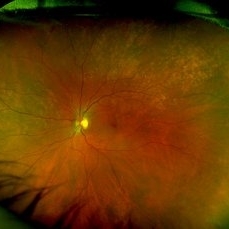 Rubella retinopathy
Jan 8 2022 by Parnian Arjmand, MD, MSc, FRCSC, DABO
Bilateral pigmentary retinopathy in a young patient with a history of congenital rubella.
Condition/keywords: bilateral pigmentary retinopathy, congenital rubella, rubella
-
 Mellaril Toxicity
Apr 2 2019 by Gary R. Cook, MD, FACS
White male with a pigmentary retinopathy OS secondary to long-term treatment with Mellaril for psychosis.
Imaging device: Topcon VT-50
Condition/keywords: bilateral pigmentary retinopathy, mellaril toxicity, secondary pigmentary degeneration
-
 Mellaril Toxicity
Apr 2 2019 by Gary R. Cook, MD, FACS
White male with a pigmentary retinopathy OD secondary to long-term treatment with Mellaril for psychosis.
Imaging device: Topcon VT-50
Condition/keywords: bilateral pigmentary retinopathy, mellaril toxicity, secondary pigmentary degeneration
-
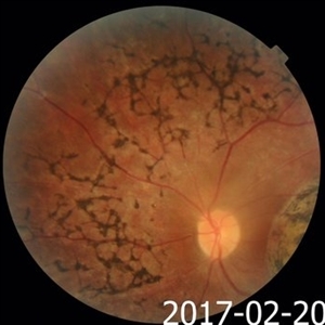
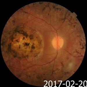 Macular Coloboma and Pigmentary Retinopathy
Feb 25 2017 by Hamid Ahmadieh, MD
Color fundus photograph of the right eye of a 25-year-old woman with the history of low vision since childhood. Bilateral macular colobomata and pigmentary retinopathy similar to Leber's congenital amaurosis are present.
Photographer: Shabnam Poureh, Negah Eye Center, Tehran, Iran
Condition/keywords: bilateral pigmentary retinopathy, color fundus photograph, macular coloboma, pigmentary retinal dystrophy
-
 Macular Coloboma and Pigmentary Retinopathy
Feb 25 2017 by Hamid Ahmadieh, MD
Color fundus photograph of the right eye of a 25-year-old woman with the history of low vision since childhood. Bilateral macular colobomata and pigmentary retinopathy similar to Leber's congenital amaurosis are present.
Photographer: Shabnam Poureh, Negah Eye Center, Tehran, Iran
Condition/keywords: bilateral pigmentary retinopathy, color fundus photograph, macular coloboma
-
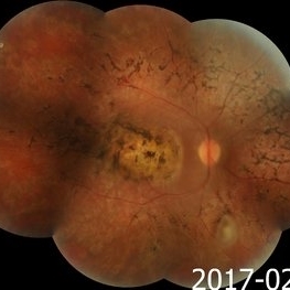 Macular Coloboma and Pigmentary Retinopathy
Feb 25 2017 by Hamid Ahmadieh, MD
Merged color fundus photograph of the right eye of a 25-year-old woman with the history of low vision since childhood. Bilateral macular colobomata and pigmentary retinopathy similar to Leber's congenital amaurosis are present.
Photographer: Shabnam Poureh, Negah Eye Center, Tehran, Iran
Condition/keywords: bilateral pigmentary retinopathy, color fundus photograph, macular coloboma, pigmentary retinal dystrophy
-
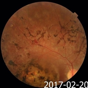
 Macular Coloboma and Pigmentary Retinopathy
Feb 25 2017 by Hamid Ahmadieh, MD
Color fundus photograph of the left eye of a 25-year-old woman with the history of low vision since childhood. Bilateral macular colobomata and pigmentary retinopathy similar to Leber's congenital amaurosis are present.
Photographer: Shabnam Poureh, Negah Eye Center, Tehran, Iran
Condition/keywords: bilateral pigmentary retinopathy, color fundus photograph, macular coloboma
-
 Macular Coloboma and Pigmentary Retinopathy
Feb 25 2017 by Hamid Ahmadieh, MD
Color fundus photograph of the left eye of a 25-year-old woman with the history of low vision since childhood. Bilateral macular colobomata and pigmentary retinopathy similar to Leber's congenital amaurosis are present.
Photographer: Shabnam Poureh, Negah Eye Center, Tehran, Iran
Condition/keywords: bilateral pigmentary retinopathy, color fundus photograph, macular coloboma
-
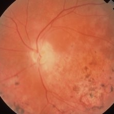
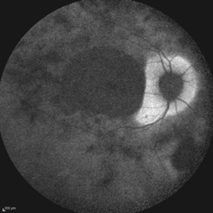
 Leber's Congenital Amaurosis
Feb 25 2017 by Hamid Ahmadieh, MD
Infrared image of the right eye of a 25-year-old woman with bilateral macular colobomata and pigmentary retinopathy similar to Leber's congenital amaurosis.
Photographer: Shabnam Poureh, Negah Eye Center, Tehran, Iran
Condition/keywords: bilateral pigmentary retinopathy, infrared image, macular coloboma
-
 Macular Coloboma and Pigmentary Retinopathy
Feb 25 2017 by Hamid Ahmadieh, MD
Fundus autofluorescence (FAF) image of the right eye of a 25-year-old woman with bilateral macular colobomata and pigmentary retinopathy similar to Leber's congenital amaurosis.
Photographer: Shabnam Poureh, Negah Eye Center, Tehran, Iran
Condition/keywords: bilateral pigmentary retinopathy, fundus autofluorescence (FAF), macular coloboma
-
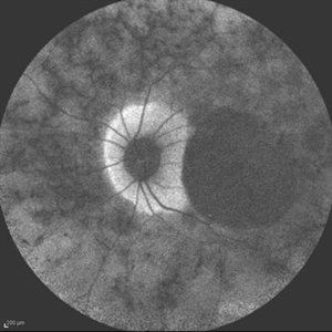
 Macular Coloboma and Pigmentary Retinopathy
Feb 25 2017 by Hamid Ahmadieh, MD
Infrared image of the left eye of a 25-year-old woman with bilateral macular colobomata and pigmentary retinopathy similar to Leber's congenital amaurosis.
Photographer: Shabnam Poureh, Negah Eye Center, Tehran, Iran
Condition/keywords: bilateral pigmentary retinopathy, infrared image, macular coloboma
-
 Macular Coloboma and Pigmentary Retinopathy
Feb 25 2017 by Hamid Ahmadieh, MD
Fundus autofluorescence (FAF) image of the left eye of a 25-year-old woman with bilateral macular colobomata and pigmentary retinopathy similar to Leber's congenital amaurosis.
Photographer: Shabnam Poureh, Negah Eye Center, Tehran, Iran
Condition/keywords: bilateral pigmentary retinopathy, fundus autofluorescence (FAF), macular coloboma
-
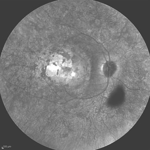
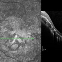 Macular Coloboma and Pigmentary Retinopathy
Feb 25 2017 by Hamid Ahmadieh, MD
Infrared and OCT images of the left eye of a 25-year-old woman with bilateral macular colobomata and pigmentary retinopathy similar to Leber's congenital amaurosis.
Photographer: Shabnam Poureh, Negah Eye Center, Tehran, Iran
Condition/keywords: bilateral pigmentary retinopathy, infrared image, macular coloboma, optical coherence tomography (OCT)
-
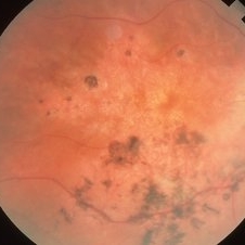
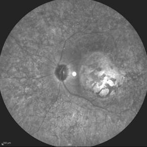
 Retinitis Pigmentosa
Aug 25 2015 by René Hernán Parada Vásquez
Fundus photograph of both eyes of a 38-year-old female with retinitis pigmentosa, bone spicule-shaped pigment deposits are present in the mid periphery, and macula with a peripheral ring of depigmentation.
Photographer: Parada René, ESO, Guatemala.
Imaging device: Canon CR-2
Condition/keywords: bilateral pigmentary retinopathy, retinitis pigmentosa, retinitis pigmentosa (RP) dystrophy
-
 Kearns-Sayre Syndrome
Sep 18 2012 by Michael P. Kelly, FOPS
Retinal fundus photograph of a Kearns-Sayre Syndrome patient.
Photographer: Michael P. Kelly, FOPS Director, Duke Eye Labs, Duke University Hospital, Duke Eye Center
Imaging device: Canon 60UV
Condition/keywords: bilateral pigmentary retinopathy, cardiac conduction abnormalities, chronic progressive ophthalmoplegia, heart-block, Kearns-Sayre Syndrome, ptosis
 Loading…