Search results (47 results)
-
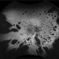
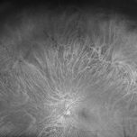 Bear Tracks CHRPE - Red Channel
Jul 29 2025 by Drew Mitchell
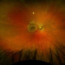
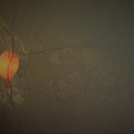
Green Free UWF image of extensive bear track patterned CHRPE.
Photographer: Drew Mitchell, OCT-C
Imaging device: Optos California
Condition/keywords: bear tracks, CHRPE, congenital hypertrophy of the retinal pigment epithelium (CHRPE), Green Free, OPTOS CALIFORNIA
-
 CHRPE Spots
Jun 13 2025 by Brandon I Fram, MD, BS
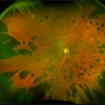
21 year-old, no past medical or family histories, with diffuse CHRPE spots suspicious for Gardner syndrome/FAP
Condition/keywords: bear tracks, CHRPE, congenital hypertrophy of the retinal pigment epithelium (CHRPE), familial adenomatous polyposis, Gardner Syndrome
-
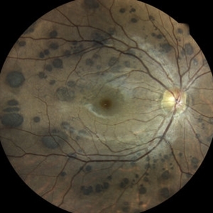
 Bear Tracks (CHRPE)
Jun 4 2025 by Paulina Araujo
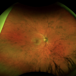
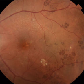
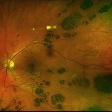
The 55-degree fundus photograph of the left eye shows bear tracks along the inferior temporal arcade.
Photographer: Paulina D.Araujo Martínez, Asociación para Evitar la Ceguera en México I.A.P., Hospital Dr Luis Sánchez Bulnes.
Condition/keywords: bear tracks, congenital hypertrophy of the retinal pigment epithelium (CHRPE)
-
 CHRPE and Bear Tracks
Jan 7 2025 by Drew Mitchell
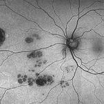
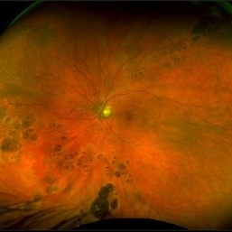
Fundus Autofluorescence of a CHRPE and Bear Tracks.
Photographer: Drew Mitchel, OCT-C
Imaging device: Optos Silverstone
Condition/keywords: bear tracks, CHRPE, congenital hypertrophy of the retinal pigment epithelium (CHRPE)
-
 CHRPE
CHRPE
Jan 6 2025 by Kavitha Duraipandi, MD DNB FICO FRCS
Bear tracks (animal tracks, grouped pigmentation spots) are simply many small CHRPEs located in isolated small area of the retina. These have not been reported to have the potential to transform into adenocarcinoma but yearly evaluations may be prudent.
Condition/keywords: CHRPE
-
 Bear Track Lesions in a Case of Congenital Hypertrophy of RPE
Sep 6 2024 by Giriraj Vibhute
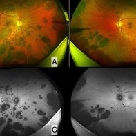
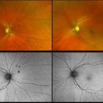
Fundus photo and autofluorescence image in a 58-year-old woman A&B: Right and left eye ultrawidefield pseudocolor imaging in case of congenital hypertrophy of RPE. C&D: Fundus autofluorescence of right and left eye of the same patient. The patient/ family members did not have any history of colon cancer. Patient was advised colonoscopy and family members were screened.
Photographer: Giriraj Vibhute, dept of retina, M M Joshi eye institute, Hubli. India
Imaging device: Optos daytona
Condition/keywords: bear tracks, congenital hypertrophy of the retinal pigment epithelium (CHRPE)
-
 CHRPE - "BEAR TRACKS" PATTERN
Nov 8 2022 by Heitor Nogueira
BEAR TRACKS
Photographer: Heitor Nogueira
Condition/keywords: bear tracks, CHRPE, congenital hypertrophy of the retinal pigment epithelium (CHRPE)
-
 Congenital Hypertrophy of RPE: "Bear Tracks"
Aug 5 2021 by Niloofar Piri, MD
Ultrawide field fundus photograph of a 79-year-old patient who was incidentally found to have extensive bear track lesions in both eyes. Left eye was treated for NVG in the past and bear tracks were only visible temporal to the macula where there was no laser scars. He was referred to be seen by gastroenterologist and have a colonoscopy given high association with FAP and Gardner's syndrome.
Photographer: Jacob Grodsky, MD, St. Louis University
Condition/keywords: bear tracks, congenital hypertrophy of the retinal pigment epithelium (CHRPE)
-
 Bear Tracks
May 14 2021 by Ronald Coriasso
Bear tracks
Photographer: Ronald Coriasso, Retina Specialist of Michigan
Condition/keywords: bear tracks
-
 Bear Tracks
May 14 2021 by Ronald Coriasso
Bear tracks
Photographer: Ronald Coriasso, Retina Specialist of Michigan
Condition/keywords: bear tracks
-
 Bear Track CHRPE OS
Jan 15 2021 by Brad Lovelace
Fundus photograph of a 24-year-old male with congenital retina pigment epithelial hypertrophy (bear tracks) OS.
Photographer: Brad Lovelace, COT, Retina Consultants of Southern Colorado, Colorado Springs
Imaging device: Optos California
Condition/keywords: bear tracks, congenital hypertrophy of the retinal pigment epithelium (CHRPE)
-
 Bear Tracks
Nov 10 2020 by Ronald Coriasso
Fundus photo of 68-year-old female with history of plaquenil use. Her findings are most consistent with bear tracks, however these kinds of lesions can be indicative of familial adenomatous polyposis (FAP).
Photographer: Ronald Coriasso
Imaging device: OPTOS
Condition/keywords: bear tracks, familial adenomatous polyposis
-
 Amelanotic Bear Tracks of the Retina
Jan 13 2020 by Sophia El Hamichi, MD
Fundus photograph of a 5-year-old patient with amelanotic bear tracks of the retina OD. No family history of colon cancer reported.
Photographer: Abby Orcutt-Hayes, Murray Ocular Oncology and Retina
Condition/keywords: amelanotic, bear tracks, congenital hypertrophy of the retinal pigment epithelium (CHRPE), pediatic retina
-
 Congenital Hypertrophy of the Retinal Pigment Epithelium
Nov 11 2019 by Jessica Norkus
Bilateral Optos ultra wide field imaging of a 31-year-old female patient with CHRPE lesions. Lesions in OD were suspicious of Gardner Syndrome due to familial history of cancerous polyps in colon. Patient underwent colonoscopy and was deemed clear.
Photographer: Jessica Norkus, COA, Retina Specialists of Michigan
Imaging device: Optos Ultra Wide Field Camera
Condition/keywords: bear tracks, bilateral, color fundus photograph, color photo, congenital hypertrophy of the retinal pigment epithelium (CHRPE), fundus autofluorescence (FAF), fundus photograph, lacunae, macula, optic disc, Optos, pseudocolor, ultra-wide field imaging
-
 Congenital Hypertrophy of Retinal Pigment Epithelium
Sep 7 2019 by Hashim Ali Khan, OD, FAAO
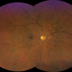
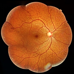
Color fundus montage of a 22-year-old man with congenital hypertrophy of retinal pigment epithelium.
Condition/keywords: bear tracks, congenital hypertrophy of the retinal pigment epithelium (CHRPE), RPE hyperplasia
-
 Congenital Hypertrophy of Retinal Pigment Epithelium (CHRPE)
Sep 7 2019 by Hashim Ali Khan, OD, FAAO
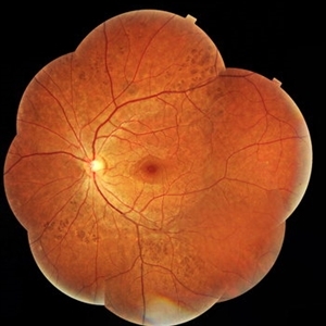
Color fundus montage of a 22-year-old man with congenital hypertrophy of retinal pigment epithelium..
Condition/keywords: bear tracks, congenital hypertrophy of the retinal pigment epithelium (CHRPE)
-
 Congenital Hypertrophy of Retinal Pigment Epithelium (CHRPE)
Sep 6 2019 by Hashim Ali Khan, OD, FAAO
Color fundus montage of a 22-year-old man with congenital hypertrophy of retinal pigment epithelium.
Imaging device: TOPCON TRC NW8F
Condition/keywords: bear tracks, congenital hypertrophy of the retinal pigment epithelium (CHRPE), RPEH-FAP
-
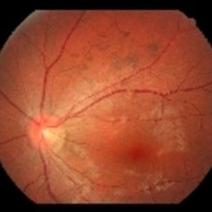
 Acute Compressive Optic Neuropathy
Jun 1 2019 by John S. King, MD
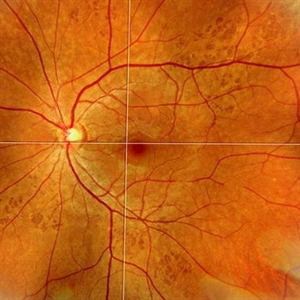
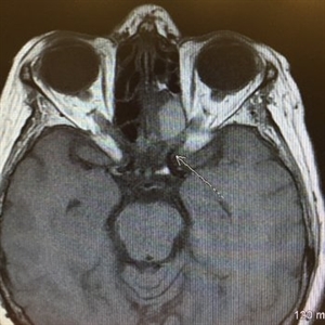
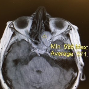
84-year-old white female with acute loss of vision in the left eye one day ago was sent here after going to the ED per primary eye provider. She described vision loss as a grey curtain that became total darkness. She had left sided temporal tenderness and some left sided neck pain. In the ED the cardiac work-up was u/r, the ESR and CRP were normal, and the CTH showed some non-specific opacification in the L ethmoid sinus. Acuity was HM OS with RAPD, normal EOMs, no proptosis or ptosis, posteriorly no SVPs were noted; the optic discs were pink and flat; no emboli or retinal whitening present; some bear tracks located nasally (see photo). She was referred to Dr. Doyle, who ordered an MRI, which showed a large mucocele with bony erosion into the left orbit, along with some ON enhancement possibly from compression (see images). She was operated that night and later recovered to 20/40 in that eye with a residual, inferior arcuate scotoma.
Condition/keywords: bear tracks, optic neuropathy
-
 Acute Optic Neuropathy Due to Large Mucocele
Jun 1 2019 by John S. King, MD
84-year-old white female with acute loss of vision in the left eye one day ago was sent here after going to the ED per primary eye provider. She described vision loss as a grey curtain that became total darkness. She had left sided temporal tenderness and some left sided neck pain. In the ED the cardiac work-up was u/r, the ESR and CRP were normal, and the CTH showed some non-specific opacification in the L ethmoid sinus. Acuity was HM OS with RAPD, normal EOMs, no proptosis or ptosis, posteriorly no SVPs were noted; the optic discs were pink and flat; no emboli or retinal whitening present; some bear tracks located nasally (see photo). She was referred to Dr. Doyle, who ordered an MRI, which showed a large mucocele with bony erosion into the left orbit, along with some ON enhancement possibly from compression (see Images). She was operated that night and later recovered to 20/40 in that eye with a residual, inferior arcuate scotoma.
Condition/keywords: bear tracks, optic neuropathy
-
 Acute Optic Neuropathy Due to Large Mucocele (Incidental Bear Tracks)
Jun 1 2019 by John S. King, MD
84-year-old white female with acute loss of vision in the left eye one day ago was sent here after going to the ED per primary eye provider. She described vision loss as a grey curtain that became total darkness. She had left sided temporal tenderness and some left sided neck pain. In the ED the cardiac work-up was u/r, the ESR and CRP were normal, and the CTH showed some non-specific opacification in the L ethmoid sinus. Acuity was HM OS with RAPD, normal EOMs, no proptosis or ptosis, posteriorly no SVPs were noted; the optic discs were pink and flat; no emboli or retinal whitening present; some bear tracks located nasally (see photo). She was referred to Dr. Doyle, who ordered an MRI, which showed a large mucocele with bony erosion into the left orbit, along with some ON enhancement possibly from compression (see images). She was operated that night and later recovered to 20/40 in that eye with a residual, inferior arcuate scotoma.
Photographer: Karin Aletter
Imaging device: Topcon 50
Condition/keywords: bear tracks, optic neuropathy
-
 Bear Tracks - Smartphone Fundus Image
Dec 27 2018 by Prithvi Chandrakanth
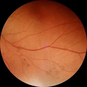
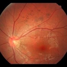
Male 43-years-old, BCVA BE 6/6, asymptomatic, Fundus RE multiple hyperpigmented circular/oval well defined lesions present inferonasally to the optic disc.
Photographer: Dr.Prithvi Chandrakanth, Dr.Chandrakanth Malabar Nethralaya, Kozhikode
Imaging device: Trash To Treasure Retcam - Smartphone Fundus Camers
Condition/keywords: bear tracks, congenital hypertrophy of the retinal pigment epithelium (CHRPE), retcam, smartphone fundus photography
-
 Bear Tracks
Apr 28 2018 by xiangbin kong
Fundus photograph of an 24-year-old woman with bear tracks.
Photographer: Yawei Dong
Imaging device: Topcon
Condition/keywords: bear tracks
-
 Bear Tracks
Apr 28 2018 by xiangbin kong
Fundus photograph of an 24-year-old woman with bear tracks.
Photographer: Yawei Dong
Imaging device: Topcon
Condition/keywords: bear tracks
-
 Bear Tracks
Apr 27 2018 by Giselle DeOliveira
Fundus Montage photograph of 13-year-old girl.
Photographer: Giselle DeOliveira, University of Miami , Bascom Palmer Eye Institute
Condition/keywords: bear tracks
-
 Bear Tracks, CHRPE
Oct 12 2017 by Theodore Leng, MD, MS, FASRS
Bear Tracks, CHRPE
Condition/keywords: bear tracks, congenital hypertrophy of the retinal pigment epithelium (CHRPE)
 Loading…