Search results (129 results)
-
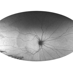
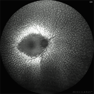
 Autofluorescence in Multiple Choroidal Ruptures
Jun 26 2025 by Hector Gabriel Moreno Solano, MD, MHA
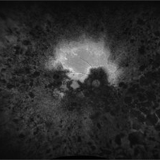
Fundus autofluorescence imaging of the right eye shows three hypoautofluorescent linear lesions located temporally to the fovea, consistent with choroidal ruptures. The lesions demonstrate sharply demarcated borders with variable surrounding hyperautofluorescence, suggestive of retinal pigment epithelium (RPE) disruption and potential remodeling. One rupture is located near the foveal region, though the foveal center remains spared.
Photographer: Hector Gabriel Moreno Solano, Instituto Mexicano de Oftalmología “IMO I.A.P”
Imaging device: CLARUS
Condition/keywords: autofluorescence imaging, Choroidal Rupture
-
 Retinitis Pigmentosa
Apr 17 2025 by Virginia Gebhart
Fundus autofluorescence of 75 year old female with Retinitis Pigmentosa. Pt diagnosed at age 53. Diffuse RPE atrophy with minimal central sparing present in both eyes. Stable and unchanged compared to previous exams. BCVA 20/200 OD, NLP OS
Photographer: Virginia Gebhart, Retina Consultants of Carolina
Imaging device: Optos California
Condition/keywords: autofluorescence imaging, bone spicule, retinitis pigmentosa, RP
-
 LCA type 10
Apr 10 2025 by Joshua Friedman
LCA type 10 due to mutations in CEP290. 36-year-old male with best corrected visual acuity of light perception in both eyes since childhood. On color fundus imaging, there is a mix of polymorphous white flecks and pigmentary changes. On autofluorescence imaging, there is almost complete loss of macular RPE. On OCT, there is complete loss of inner and outer retinal layers, the greatest losses occurring centrally.
Photographer: Stephen Tsang, MD, PhD
Condition/keywords: Leber Congenital Amaurosis
-
 Toxic Maculopathy (Elmiron)
Apr 9 2025 by Virginia Gebhart
79 year old male with toxic maculopathy from long term use of Elmiron (15+ yrs.) On exam there is stippled RPE changes, pigment clumping, and subretinal deposits. BCVA 20/100 | 20/40.
Photographer: Virginia Gebhart, Retina Consultants of Carolina
Imaging device: Optos California
Condition/keywords: autofluorescence imaging, cystoid macular degeneration, Elmiron Toxicity, Toxic Maculopathy
-
 Choroidal Melanoma with Exudative Detachment
Apr 7 2025 by Virginia Gebhart
Autofluorescence image of 36 year old female showing demarcation line of fluid/detachment from new choroidal melanoma. Pt will be scheduled for brachytherapy pending CT scan results.
Photographer: Virginia Gebhart, Retina Consultants of Carolina
Imaging device: Optos California
Condition/keywords: Autoflourescence, autofluorescence imaging, choroidal melanoma, melanoma, retinal detachment
-
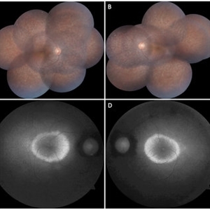
 Choroidal Hemangioma 4 Ways
Mar 13 2025 by Virginia Gebhart
Color fundus, FAF, late FA, late ICG of 64 year old male with choroidal hemangioma. Early hyperfluorescence with late leakage on FA, early hypercyanescence with late washout (25 min) on ICG.
Photographer: Virginia Gebhart, Retina Consultants of Carolina
Imaging device: Optos California
Condition/keywords: autofluorescence imaging, choroidal hemangioma, FA late phase, Fluorescein angiography, hemangioma, indocyanine green (ICG) angiography
-
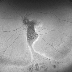
 Hemangioma of Retina (FAF)
Mar 5 2025 by Virginia Gebhart
Fundus autofluorescence of 64 year old male with choroidal hemangioma in the macula and STA. Persistent IRF and new cuff of SRF compared to previous photos. BCVA CF@face. Pt has had PDT in the past with no significant improvement. Will observe closely
Photographer: Virginia Gebhart, Retina Consultants of Carolina
Imaging device: Optos California
Condition/keywords: autofluorescence imaging, hemangioma, inferior subretinal fluid
-
 Resolved Multiple Evanescent White Dot Syndrome
Feb 18 2025 by Jordyn Beckman
Autofluorescence fundus photographs of Resolved Multiple Evanescent White Dot Syndrome in a 28 year old female with previous hyperfluorescent punctate spots throughout the posterior pole.
Photographer: Jordyn Beckman, Retina Consultants of Carolina, P.A.
Imaging device: California Optos
Condition/keywords: autofluorescence imaging, grey-white lesions, multiple evanescent white dot syndrome (MEWDS), scattered punctate
-
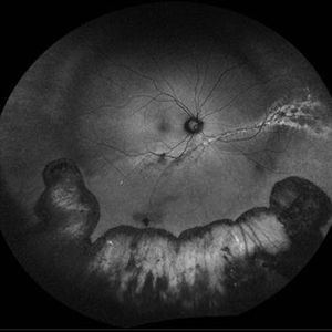
 Mac-on Retinal Detachment (Barely!)
Feb 6 2025 by Virginia Gebhart
FAF of 46 year old male with a mac-on retinal detachment from 1:00 to 6:00 with a single break at 3:00. Pt scheduled for emergent PPV/Laser/GFE
Photographer: Virginia Gebhart, Retina Consultants of Carolina
Imaging device: Optos California
Condition/keywords: autofluorescence imaging, retinal detachment
-
 Foveal Hypoplasia AF
Feb 1 2025 by Poornachandra B, MS, FVRS
This is a wide field autofluorescence image of 21 year-old male. He presented with history of low vision since childhood associated with nystagmus. Uniform fluorescence across posterior pole with absent foveal hypo autofluorescence can be seen on the image.
Photographer: Mr Dhikshith
Condition/keywords: autofluorescence imaging, foveal hypoplasia, nystagmus, ultra-wide field imaging
-
 Elmiron Toxicity
Jan 15 2025 by Virginia Gebhart
54 year old female with pigmentary degeneration secondary to Elmiron. Stippled RPE maculopathy has lightly progressed with stable vision compared to previous visits. BCVA 20/200 OU. Pt reports taking Elmiron from 2010 to 2019.
Photographer: Virginia Gebhart
Imaging device: Optos California
Condition/keywords: autofluorescence imaging, Maculopathy, secondary pigmentary degeneration
-
 Repaired Retinal Detachment with Multiple Breaks
Dec 9 2024 by Virginia Gebhart
FAF in 25 year old female of repaired retinal detachment 1.5 year s/p scleral buckle/cryo. Pt had been having symptoms for over a year, inferior demarcation line from retinal fluid that was present. Retina remains flat and attached under buckle. Treated lattice inferiorly, no new holes or tears. VA 20/20
Photographer: Virginia Gebhart, Retina Consultants of Carolina
Imaging device: Optos California
Condition/keywords: autofluorescence imaging, cryotherapy, demarcation line, lattice degeneration, scleral buckle
-
 Fundus Flavimaculatus Fundus Autofluorescence Imaging
Sep 25 2024 by Keshavi Shah
FAF imaging of a 37 year old male patient with Stargardt's Disease of adult onset ( Fundus Flavimaculatus) presenting with dimunition of night vision and dyschromatopsia demonstrating areas of hypo-auto fluorescence (representing RPE/ Photo-receptor atrophy) and hyper-autofluorescence(representing excessive lipo-fuschin accumulation in the RPE cells) with peri-papillary sparing, typical of ABCA-4 related disorders.
Photographer: Simran
Imaging device: Optos Daytona
Condition/keywords: fundus autofluorescence (FAF), fundus flavimaculatus
-
 Fundus Flavimaculatus Fundus Autofluorescence Imaging
Sep 25 2024 by Keshavi Shah
FAF imaging of a 37 year old male patient with Stargardt's Disease of adult onset ( Fundus Flavimaculatus) presenting with dimunition of night vision and dyschromatopsia demonstrating areas of hypo-auto fluorescence (representing RPE/ Photo-receptor atrophy) and hyper-autofluorescence(representing excessive lipo-fuschin accumulation in the RPE cells) with peri-papillary sparing, typical of ABCA-4 related disorders.
Photographer: Simran
Imaging device: Nikon Optos Daytona
Condition/keywords: fundus autofluorescence (FAF), fundus flavimaculatus
-
 Serpiginous Choroidopathy Autofluorescence
Sep 24 2024 by Gustavo Uriel Fonseca Aguirre
Autofluorescence image of the right fundus of a 32-year-old female patient diagnosed with serpiginous choroiditis.
Photographer: Gustavo U. Fonseca Aguirre, Fundación Hospital Nuestra Señora de la Luz, Ciudad de México
Condition/keywords: autofluorescence imaging, serpiginous choroiditis
-
 Robson-Holder Ring
Jul 15 2024 by Arthi Mohankumar , MS,MRCS ED, FICO,FAICO
Robson holder hyper autofluorescent ring in a patient with retinitis pigmentosa.
Photographer: Arthi Mohankumar
Condition/keywords: autofluorescence imaging, retinitis pigmentosa (RP) dystrophy, Rod cone dystrophy
-
 Central Serous Chorioretinopathy With Comet Tail Sign
Jul 13 2024 by Aditya S Kelkar, MS, FRCS, FASRS,FRCOphth
Fundus Autofluorescence imaging of an 59-year-old man with Chronic Central Serous Chorioretinopathy demonstrating a Comet Tail Sign.
Photographer: Dr. Rabia Naaz, National institute of Ophthalmology, Pune, India
Imaging device: OPTOS DAYTONA
Condition/keywords: central serous chorioretinopathy (CSCR)
-
 FAF of Barricade Laser on Choroidal Osteoma
Jun 12 2024 by Virginia Gebhart
20 year old female with stable choroidal osteoma s/p PDT x 3 and focal laser x 2. No obvious progression on last exam, vision 20/30. Monitoring closely.
Photographer: Virginia Gebhart
Imaging device: Topcon 50 DX
Condition/keywords: autofluorescence imaging, barrier laser, choroidal osteoma, focal laser, fundus autofluorescence (FAF)
-
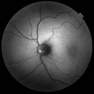 Autofluorescence in Optic Nerve Head Drusen
May 28 2024 by Nishikant J Borse, MS, FMRF, FASRS
65-year-old female was referred for disc edema. An Autofluorescence Imaging was done which showed the autofluorescence of the optic nerve head drusen.
Photographer: Dr Nishikant Borse , Insight eye Clinic , Mumbai
Imaging device: Topcon Triton
Condition/keywords: Autofluorescence imaging of Optic Disc Drusen
-
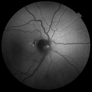 Autofluorescence in Optic Nerve Head Drusen
May 28 2024 by Nishikant J Borse, MS, FMRF, FASRS
65-year-old female was referred for disc edema. An Autofluorescence Imaging was done which showed the autofluorescence of the optic nerve head drusen.
Photographer: Dr Nishikant Borse , Insight eye Clinic , Mumbai
Imaging device: Topcon Triton
Condition/keywords: Autofluorescence imaging of Optic Disc Drusen
-
 Central Serous Retinopathy
Mar 19 2024 by Corey Grant
Ultra Wide-Field Fundus Autofluorescence Imaging of a 37 year old female with Central Serous Retinopathy affecting her right eye. Patient Visual Acuity was 20/20 in both eyes. Patient reported black spots in her vision onset three years ago, with associating flashes of light. Patient reports history of cortisone back injections a few years ago and denies Flonase use. The physician stated that there is hyperautofluorescence in the area of gutter of Sub-Retinal Fluid which likely happened from CSR.
Photographer: Corey Grant, OSC
Imaging device: OPTOS CALIFORNIA RGB
Condition/keywords: Central Serous Chorioretinopathy (CSR), central serous retinopathy (CSR), fundus autofluorescence (FAF), Guttering, hyperautofluorescence, inferior retina, OPTOS, Retina, Right Eye, subretinal fluid, ULTRA WIDE FIELD
-
 Acute Posterior Multifocal Placoid Pigment Epitheliopathy
Feb 20 2024 by Soobien Lee
Optos fundus autofluorescence photograph of a 20-year-old caucasian female with viral prodrome and vision loss OS>OD secondary to Acute Posterior Multifocal Placoid Pigment Epitheliopathy (APPME). Imaging of her left eye shows hypoautofluorescent areas corresponding to multiple bilateral placoid lesions at the level of RPE and choroid throughout the posterior pole.
Photographer: Ashley Metzger, Elman Retina Group
Imaging device: Optos Ultra-Widefield Autoflurescence Imaging
Condition/keywords: acute posterior multifocal placoid pigment epitheliopathy (APMPPE), autofluorescence imaging, bacilliary layer detachment, Optos, OPTOS CALIFORNIA, uveitis, white dot syndrome
-
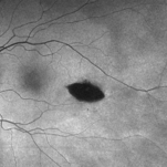 Torpedo Maculopathy
Feb 20 2024 by Soobien Lee
Optos fundus autofluorescence photograph of a 35-year-old asymptomatic female with no ocular or medical history with stable and chronic appearing torpedo-shaped macula lesion in the left eye.
Photographer: Peter Sotirakos, Elman Retina Group
Imaging device: Optos Ultra-Widefield Autoflurescence Imaging
Condition/keywords: autofluorescence imaging, genetics, macula, maculopathy, Optos, torpedo maculopathy
-
 Benign Familial Fleck Retina
Dec 21 2023 by Vishal Agrawal, MD, FRCS,FACS,FASRS
Green Autoflourescence image of fleck retinopathy.
Photographer: Dr Ayushi
Imaging device: Clarus 700
Condition/keywords: autofluorescence imaging, fleck retinopathy
-
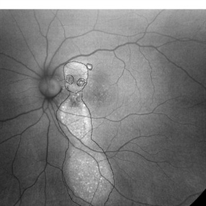 Chronic CSR - Dancing doll
Nov 20 2023 by Harsh Vardhan Singh, MS
37-year male with chronic CSR
Photographer: Harsh Vardhan Singh
Imaging device: Zeiss clarus 700
Condition/keywords: autofluorescence imaging, Central Serous Chorioretinopathy (CSR), fundus autofluorescence (FAF), idiopathic central serous choroidopathy (ICSC)
 Loading…