Search results (266 results)
-
 Acute Posterior Multifocal Placoid Pigment Epitheliopathy
Feb 20 2024 by Soobien Lee
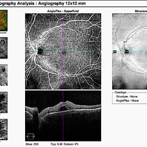
12x12mm OCT Angiography of a 20-year-old caucasian female with viral prodrome and vision loss OS>OD secondary to Acute Posterior Multifocal Placoid Pigment Epitheliopathy (APPME). Imaging shows multifocal flow voids.
Photographer: Kim Seay, Elman Retina Group
Imaging device: 12x12mm OCT-Angiography
Condition/keywords: acute posterior multifocal placoid pigment epitheliopathy (APMPPE), bacillary layer detachment, OCT, OCT Angiography, Uveitis, white dot syndrome
-
 Acute Posterior Multifocal Placoid Pigment Epitheliopathy
Feb 20 2024 by Soobien Lee
A 20-year-old caucasian female with viral prodrome and vision loss OS>OD secondary to Acute Posterior Multifocal Placoid Pigment Epitheliopathy (APPME). OCT of the left macula shows bacillary layer detachment.
Photographer: Kim Seay, Elman Retina Group
Condition/keywords: acute posterior multifocal placoid pigment epitheliopathy (APMPPE), bacilliary layer detachment, OCT, Uveitis, white dot syndrome
-
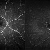
 Acute Posterior Multifocal Placoid Pigment Epitheliopathy
Feb 20 2024 by Soobien Lee
Fluorescein angiogram of a 20-year-old caucasian female with viral prodrome and vision loss OS>OD secondary to Acute Posterior Multifocal Placoid Pigment Epitheliopathy (APPME). Early blockage with late hyperfluorescent leakage can be seen on fluorescein angiography of the left eye.
Photographer: Ashley Metzger, Elman Retina Group
Imaging device: Optos Ultra-Widefield Fluorescein Angiography
Condition/keywords: acute posterior multifocal placoid pigment epitheliopathy (APMPPE), bacilliary layer detachment, FA, FA late phase, FA late phase leakage, fluorescein angiogram (FA), Optos, uveitis, white dot syndrome
-
 Acute Posterior Multifocal Placoid Pigment Epitheliopathy
Feb 20 2024 by Soobien Lee
Fluorescein angiogram of a 20-year-old caucasian female with viral prodrome and vision loss OS>OD secondary to Acute Posterior Multifocal Placoid Pigment Epitheliopathy (APPME). Early blockage with late hyperfluorescent leakage can be seen on fluorescein angiography of the left eye.
Photographer: Ashley Metzger, Elman Retina Group
Imaging device: Optos Ultra-Widefield Fluorescein Angiography
Condition/keywords: acute posterior multifocal placoid pigment epitheliopathy (APMPPE), bacilliary layer detachment, FA, FA early phase, fluorescein angiogram (FA), Optos, uveitis, white dot syndrome
-
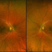
 Acute Posterior Multifocal Placoid Pigment Epitheliopathy
Feb 20 2024 by Soobien Lee
Optos fundus autofluorescence photograph of a 20-year-old caucasian female with viral prodrome and vision loss OS>OD secondary to Acute Posterior Multifocal Placoid Pigment Epitheliopathy (APPME). Imaging of her left eye shows hypoautofluorescent areas corresponding to multiple bilateral placoid lesions at the level of RPE and choroid throughout the posterior pole.
Photographer: Ashley Metzger, Elman Retina Group
Imaging device: Optos Ultra-Widefield Autoflurescence Imaging
Condition/keywords: acute posterior multifocal placoid pigment epitheliopathy (APMPPE), autofluorescence imaging, bacilliary layer detachment, Optos, OPTOS CALIFORNIA, uveitis, white dot syndrome
-
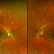
 Acute Posterior Multifocal Placoid Pigment Epitheliopathy
Feb 20 2024 by Soobien Lee
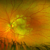
Optos color fundus photograph of a 20-year-old caucasian female with viral prodrome and vision loss OS>OD secondary to Acute Posterior Multifocal Placoid Pigment Epitheliopathy (APPME). Imaging of her left eye shows multiple bilateral creamy yellow-white placoid lesions at the level of RPE and choroid throughout the posterior pole.
Photographer: Ashley Metzger, Elman Retina Group
Imaging device: Optos Ultra-Widefield Imaging
Condition/keywords: acute posterior multifocal placoid pigment epitheliopathy (APMPPE), bacilliary layer detachment, Optos, uveitis, white dot syndrome
-
 acute posterior multifocal placoid pigment epitheliopathy
Sep 23 2022 by Jaideep sharma
A 50-year old woman presented to us with unilateral progressive and painless visual blurring. She was diagnosed as a case of CSCR and started on topical dorzolamide with no improvement in VA. Her best-corrected visual acuity (BCVA) was RE 6/6 and LE 6/60 . Eye examination revealed vitritis (grade1) with optic disc hyperemia and multiple serous retinal detachments with choroidal striae in the left eye and a normal right eye. She is k/c/o diabetes. Her past ocular and drug histories were unremarkable. Retinal imaging revealed characteristic features of APMPPE in the left eye. All laboratory testing results were inconclusive. VA and OCT findings significantly improved following the treatment with LE posterior sub tenon’s triamcinolone (40 mg/ml). 1 month post injection VA of the left eye reached 6/6 with resolved serous retinal detachments in this eye. This case is unique as it was managed via PST injection rather than conventional steroid therapy
Photographer: jaideep sharma jaipur calgary eye hospital rajasthan india
Condition/keywords: acute posterior multifocal placoid pigment epitheliopathy (APMPPE), FFA
-
 Acute posterior multifocal placoid pigment epitheliopathy (APMPPE)
Sep 23 2022 by Jaideep sharma
A 50-year old woman presented to us with unilateral progressive and painless visual blurring. She was diagnosed as a case of CSCR and started on topical dorzolamide with no improvement in VA. Her best-corrected visual acuity (BCVA) was RE 6/6 and LE 6/60 . Eye examination revealed vitritis (grade1) with optic disc hyperemia and multiple serous retinal detachments with choroidal striae in the left eye and a normal right eye. She is k/c/o diabetes. Her past ocular and drug histories were unremarkable. Retinal imaging revealed characteristic features of APMPPE in the left eye. All laboratory testing results were inconclusive. VA and OCT findings significantly improved following the treatment with LE posterior sub tenon’s triamcinolone (40 mg/ml). 1 month post injection VA of the left eye reached 6/6 with resolved serous retinal detachments in this eye. This case is unique as it was managed via PST injection rather than conventional steroid therapy
Photographer: jaideep sharma jaipur calgary eye hospital rajasthan india
Condition/keywords: acute posterior multifocal placoid pigment epitheliopathy (APMPPE), FFA
-
 Acute Posterior Multifocal Placoid Pigment Epitheliopathy (APMPPE)
Oct 8 2020 by Mihir Trivedi
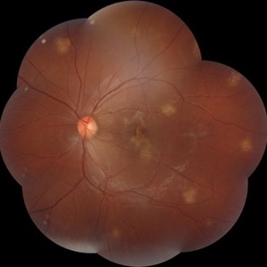
Fundus photograph of a 21-year-old male having complaints of blurring of vision. Had a viral prodrome 2 weeks before the onset of symptoms.
Imaging device: DRI OCT-1 Triton (plus) TOPCON
Condition/keywords: acute multifocal placoid pigment epitheliopathy (AMPPE)
-
 Serpiginous Choroiditis (Recurrent)
Sep 22 2019 by Haider Ali
35-year-old female presented with decrease in vision in her left eye for last 4 days and in right eye for last 8 days. Her right eye was previously involved in a similar episode about 5-6 months ago for which she was treated with oral steroids.
Photographer: Dr Haider Ali Chaudhry, Madinah Teaching Hospital, Faisalabad
Condition/keywords: acute posterior multifocal placoid pigment epitheliopathy (APMPPE), macula serpiginous choroidopathy, serpiginous choroiditis, white dot syndrome
-
 Serpiginous Choroiditis
Sep 22 2019 by Haider Ali
35-year-old female presented with decrease in vision in her left eye for last 4 days and in right eye for last 8 days. Her right eye was previously involved in a similar episode about 5-6 months ago for which she was treated with oral steroids.
Photographer: Dr Haider Ali Chaudhry, Madinah Teaching Hospital, Faisalabad
Condition/keywords: acute posterior multifocal placoid pigment epitheliopathy (APMPPE), macula serpiginous choroidopathy, posterior uveitis, serpiginous choroiditis, uveitis, white dot lesions, white dot syndrome
-
 Acute Posterior Multifocal Placoid Pigment Epitheliopathy
Jan 4 2019 by Cláudia Farinha
Composite image of both eyes of a 27-year-old male with APMPPE. In the fundus photograph, multiple yellowish placoid lesions are observed in the posterior pole in both eyes. The ICGA revealed more lesions than those observed in fundoscopy, and these were hypofluorescent through the angiogram as expected. The en face OCTA segmented at the level of the choriocapillaris revealed areas of ischemia in close correspondence with the hypofluorescent lesions (image superimposed in ICGA ). The OCT b-scan with superimposed flow shows disruption and hyperreflectivity of the external retinal layers in the affected areas and again the absence of flow in the choriocapillaris underneath. A systemic study was carried out to exclude other inflammatory and infectious causes of placoid retinochoroidopathy. The clinical picture resolved after approximately one month from the onset, without recurrence.
Photographer: Pedro Melo, Ophthalmology Department, Centro Hospitalar e Universitário de Coimbra, Coimbra Portugal
Condition/keywords: acute posterior multifocal placoid pigment epitheliopathy (APMPPE), white dot syndrome
-
 Acute Posterior Multifocal Placoid Pigment Epitheliopathy
May 3 2018 by Alexandr Stepanov
Acute posterior multifocal placoid pigment epitheliopathy.
Photographer: Alexandr Stepanov MD, PhD, FEBO, Faculty Hospital Hradec Kralove, Czech Republic
Condition/keywords: acute posterior multifocal placoid pigment epitheliopathy (APMPPE)
-
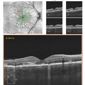
 APMPPE
Dec 14 2017 by John S. King, MD
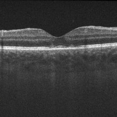
Initial scan
Imaging device: cirrus
Condition/keywords: acute posterior multifocal placoid pigment epitheliopathy (APMPPE), white dot syndrome
-
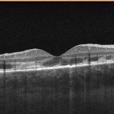
 APMPPE
Dec 14 2017 by John S. King, MD
One week later, same slice as initial scan.
Imaging device: cirrus
Condition/keywords: acute posterior multifocal placoid pigment epitheliopathy (APMPPE), white dot syndrome
-
 APMPE
APMPE
Dec 14 2017 by John S. King, MD
One week later, same slice as initial scan.
Imaging device: cirrus
Condition/keywords: acute posterior multifocal placoid pigment epitheliopathy (APMPPE), white dot syndrome
-
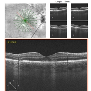 APMPPE
Dec 14 2017 by John S. King, MD
Initial scan.
Imaging device: cirrus
Condition/keywords: acute posterior multifocal placoid pigment epitheliopathy (APMPPE), white dot syndrome
-
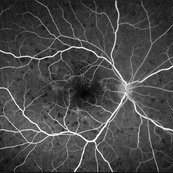 APMPPE
Dec 14 2017 by John S. King, MD
30 sec - initial visit.
Imaging device: optos
Condition/keywords: acute posterior multifocal placoid pigment epitheliopathy (APMPPE), white dot syndrome
-
 APMPPE
Dec 14 2017 by John S. King, MD
Mid and late FA - initial presentation
Imaging device: optos
Condition/keywords: acute posterior multifocal placoid pigment epitheliopathy (APMPPE), white dot syndrome
-
 APMPPE
Dec 14 2017 by John S. King, MD
About a week later
Imaging device: optos
Condition/keywords: acute posterior multifocal placoid pigment epitheliopathy (APMPPE), white dot syndrome
-
 APMPPE
Dec 14 2017 by John S. King, MD
Initial pics.
Imaging device: optos
Condition/keywords: acute posterior multifocal placoid pigment epitheliopathy (APMPPE), white dot syndrome
 Loading…