Search results (13 results)
-
 Leukemic optic nerve infiltration
Mar 31 2022 by Franco Benvenuto, MD
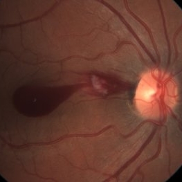
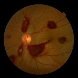
Fundus photograph of a 21-year-old otherwise healthy male presenting with acute onset of blurry vision in his both eyes for one week. Acute promyelocytic leukemia was confirmed by bone marrow study. The fundus exam showed bilateral optic nerve infiltration: Leukemic optic neuropathy.
Photographer: Franco Benvenuto, Universidad de Buenos Aires, Argentina; Universidad de Guadalajara, México
Condition/keywords: acute leukemia, infiltration of the optic nerve
-
 Choroidal MRSA Abscess
Apr 15 2021 by Rui Zhang, BA
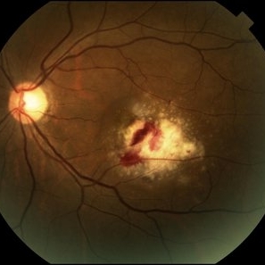
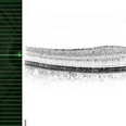
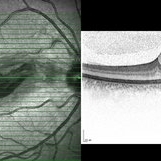
A 14-year-old boy receiving induction chemotherapy for acute lymphocytic leukemia (ALL) complained of floaters and central scotoma in his left eye. (A) Fundus photography showed sub-macular choroidal abscess with intraretinal hemorrhage and edema. (B) OCT confirmed that the abscess had not penetrated the retinal pigment epithelium (RPE). Due to systemic septic signs (fever, tachycardia, tachypnea, new-onset papules), blood cultures were drawn and they came back positive for methicillin-resistant staphylococcus aureus (MRSA). Patient was promptly treated with both IV and intravitreal antibiotics. This is a case of sub-macular choroidal MRSA abscess in the setting of systemic bacteremia in an immunocompromised host.
Photographer: Raymond Mok, CRA COMT (Dartmouth-Hitchcock Medical Center)
Imaging device: Optical coherence tomography
Condition/keywords: abscess, acute leukemia, MRSA sepsis
-
 Slide 9-103
Feb 26 2019 by Lancaster Course in Ophthalmology
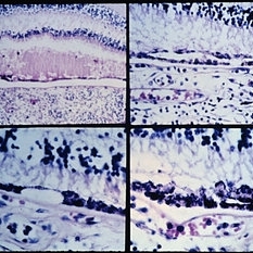
Acute leukemia with focal areas of RPE defects (lower left), and areas of double-row (upper right) and nodular (lower right) RPE hyperplasia.
Condition/keywords: acute leukemia, retinal pigment epithelium
-
 Acute Myeloid Leukemia
Dec 4 2018 by Linda A Cernichiaro- Espinosa, MD
Fundus photograph of a 12-year-old girl with superficial and deep retinal hemorrhages associated to acute myeloid leukemia (AML). A subhyaloid bleed involves the macula in both eyes.
Photographer: Dr. Linda A Cernichiaro Espinosa
Imaging device: inView (Volk Inc. USA) with iPhone 6
Condition/keywords: acute leukemia, leukemia, retinopathy, Roth spots
-
 Leukemic Retinopathy
Apr 11 2018 by Yi-Ting Hsieh, MD
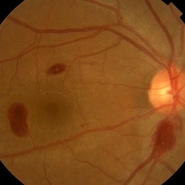
Fundus photograph of a 48-year-old otherwise healthy male presenting with acute onset of blurry vision in his both eyes for one week. Acute promyelocytic leukemia with low platelet count then was confirmed by bone marrow study.
Photographer: Chia-Chieh Hsiao, Department of Ophthalmology, National Taiwan University Hospital
Imaging device: Canon CR-DGi
Condition/keywords: acute leukemia, retinal hemorrhage
-
 Multiple Blot Hemorrhages and Roth Spots
Jan 24 2018 by Gabriel Costa Andrade, PhD
Multiple blot hemorrhages and Roth spots in a patient with acute leukemia.
Photographer: Gabriel Andrade, MD
Condition/keywords: leukemia, Roth spots
-
 CMV-Macula
Feb 24 2014 by Susanna S. Park, MD, PhD
Fundus photograph of a 59-year-old woman on chemotherapy for acute lymphocytic leukemia with new vision loss from cytomegalovirus retinitis
Photographer: Ellen Redenbo, UC Davis Eye Center
Condition/keywords: acute leukemia, CMV retinitis
-
 Leukemia 3
Mar 16 2013 by Roy Schwartz, MD
Fundus photograph of a 35-year-old man presenting with a sudden vision loss in his right eye. Macular hemorrhage with cotton wool spot were seen. Diagnostic work up revealed chronic eosinophilic leukemia.
Photographer: Galit Yair-Pur
Condition/keywords: acute leukemia, macular hemorrhage
-
 Leukemia 2
Mar 16 2013 by Roy Schwartz, MD
Six weeks after presentation, complete resolution of the hemorrhage is seen.
Photographer: Galit Yair-Pur
Condition/keywords: acute leukemia, macular hemorrhage
-
 Leukemia1
Mar 16 2013 by Roy Schwartz, MD
At presentation, spectral domain OCT shows intraretinal and sub-ILM hemorrhage as well as thickening of the RNFL in the area of the cotton wool spot.
Photographer: Galit Yair-Pur
Condition/keywords: acute leukemia, macular hemorrhage
-
 Leukemic Retinopathy
Oct 9 2012 by Sharon Fekrat, MD FACS FASRS
22-year-old female with new diagnosis of acute myelogenous leukemia. White blood cell count was 35,000,000,000 cells/L. Note Roth Spots.
Photographer: Tiffanie Keaton, Duke Eye Imaging, Durham, NC
Condition/keywords: acute leukemia, white centered retinal hemorrhage (Roth Spot)
-
 Ocular Manifestation of Acute Leukemia
Sep 8 2012 by Hamid Ahmadieh, MD
Color fundus photograph of a 26-year-old man with acute leukemia.
Photographer: Hamid Ahmadieh, MD, Ophthalmic Research Center, Labbafinejad Medical Center, Shahid Beheshti University of Medical Sciences , Tehran
Imaging device: Topcon Fundus Camera
Condition/keywords: acute leukemia, white centered retinal hemorrhage (Roth Spot)
-
 Ocular Manifestation of Acute Leukemia
Sep 5 2012 by Hamid Ahmadieh, MD
Color fundus photograph of a 26-year-old man with acute leukemia.
Photographer: Hamid Ahmadieh, MD, Ophthalmic Research Center, Labbafinejad Medical Center, Shahid Beheshti University of Medical Sciences
Imaging device: Topcon Fundus Camera
Condition/keywords: acute leukemia, white centered retinal hemorrhage (Roth Spot)
 Loading…