Search results (88 results)
-
 Pseudoxanthoma Elasticum
Dec 3 2024 by Dr Bilal Mir
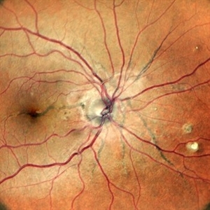
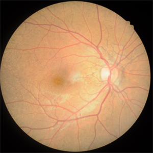
This is a fundus picture showing angiod streaks, CNVM, comet lesion.
Photographer: Dr Bilal Ahmed mir MS ophthalmology
Imaging device: Zeiss fundus camera
Condition/keywords: Angiod streaks in Pseudoxanthoma elasticum, Pseudoxanthoma elasticum
-
 Optic Disc Drusen and Angioid Streaks in Pseudoxanthoma Elasticum
Nov 19 2024 by Rafael Robbs
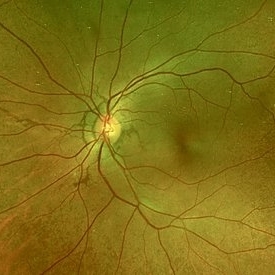
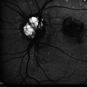
Fundus Autofluorescence: presence of optic disc drusen, associated with angioid streaks, in a patient with pseudoxanthoma elasticum.
Photographer: Rafael Robbs, Universidade Federal Fluminense, Niterói Rio de Janeiro, Brazil
Imaging device: DRI OCT-1 Triton / Triton Plus
Condition/keywords: angioid streaks, optic disc drusen, pseudoxanthoma elasticum (PXE)
-
 Angioid Streaks
Sep 29 2024 by Tejaswita Verma
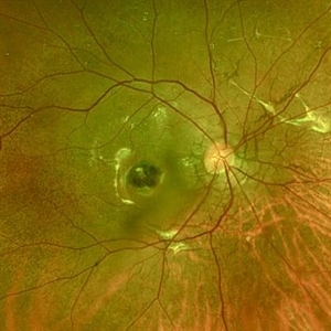
Fundus photograph of a 35 year-old female with 6/6 vision in RE , unremarkable anterior segment and family history of angioid streaks and pseudoxanthoma elasticum in sister. Fundus examination revealed angioid streaks radiating from disc , sparing the fovea .Her Sister had received multiple anti VEGF injections for angioid streaks with CNVM.
Photographer: DR. TEJASWITA VERMA
Imaging device: MIRANTE
Condition/keywords: angioid streaks
-
 Intriguing Web
Aug 28 2024 by Hemanth Murthy, MBBS, MD, FASRS
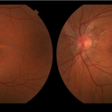
Left eye of a 43 year female patient came with blurring of vision of right eye since 2 years. There was loose redundant skin in the neck and axilla. Angiod streaks were in a spider web appearance .Vision was 1/60 in right eye and 6/9 in left eye
Photographer: Mr Veda Vyas
Imaging device: Optos Daytona
Condition/keywords: Angiod streaks in Pseudoxanthoma elasticum
-
 Intriguing Web
Aug 28 2024 by Hemanth Murthy, MBBS, MD, FASRS
Right eye of a 43 year female patient came with blurring of vision of right eye since 2 years. There was loose redundant skin in the neck and axilla. Angiod streaks were in a spider web appearance .Vision was 1/60 in right eye and 6/9 in left eye. Right macula showed a sub retinal scar with pigmentation.
Photographer: Mr Veda Vyas
Imaging device: Optos Daytona
Condition/keywords: Angiod streaks in Pseudoxanthoma elasticum
-
 Pseudoxanthoma Elasticum Associated Angioid Streaks
Aug 18 2024 by KANWALJEET HARJOT MADAN, M.S. (Ophthalmology); FAICO (Vitreous - Retina)
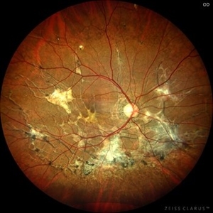
This is fundus photograph of a young 31 years male patient depicting Angioid streaks emanating from optic nerve towards the periphery and subretinal fibrosis. There is peau de orange appearance temporal to fovea with Salmon Spots in periphery. He was diagnosed to have Pseudoxanthoma Elasticum.
Photographer: Dr. Kanwaljeet Harjot Madan, M.S. (Ophthalmologist) Fellow in Vitrous & Retina. Thind Eye Hospital, Jalandhar City. Punjab. India
Imaging device: Zeiss Clarus
Condition/keywords: Angioid Streaks, fundus photograph, pseudoxanthoma elasticum (PXE)
-
 Pseudoxanthoma Elasticum with Angioid Streaks
May 10 2024 by Ethan K Sobol, MD
An asymptomatic patient with biopsy proven pseudoxanthoma elasticum. Both eyes had prominent peripapillary angioid streaks and a peau d'orange fundus appearance in the temporal macula.
Condition/keywords: angioid streaks, peau d'orange fundus, pseudoxanthoma elasticum (PXE)
-
 Angioid Streaks
Jun 14 2022 by Kingston Rodolfo Ureña-Wong, MD, Opht, MSc
Fundus photograph of an 26-year-old woman with pseudoxanthoma elasticum and angioid streaks. She developed a choroidal neovascular membrane which was treated with anti-VEGF successfully.
Photographer: Kingston Rodolfo Ureña-Wong, Asociación para evitar la ceguera en México, México.
Imaging device: Zeiss Clarus
Condition/keywords: Angioid Streaks
-
 Severe macular atrophy secondary to Pseudoxanthoma Elasticum and CNV
Jul 26 2019 by Olivia Rainey
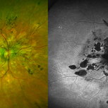
Ultra-wide field color/autofluorescence comparison of a 54-year-old female with severe macular atrophy secondary to pseudoxanthoma elasticum. Patient developed choroidal neovascularization that does not warrant treatment due to the patient's poor visual acuity. Patient has significantly attenuated ERG. Genetic testing is recommended to rule out retinitis pigmentosa. The mechanism is yet to be determined, but there is a thought that ABCC6 mutation causes alteration of plasma lipid composition may be implicated in the systemic changes observed in PXE.
Photographer: Olivia Rainey
Imaging device: Optos
Condition/keywords: angioid streaks, atrophic central lesion, autofluorescence imaging, choroidal neovascularization (CNV), hypofluorescent lesions, macular atrophy, Optos, pigmentary retinal dystrophy, pseudoxanthoma elasticum (PXE), ultra-wide field imaging
-
 Slide 9-95
Feb 26 2019 by Lancaster Course in Ophthalmology
Macular disciform lesion in a 73-year-old man with angioid streaks and pseudoxanthoma elasticum. The streak is subjacent to the scar and is the point at which choroidal vessels (arrows) extend internally. The same streak closer to the disc is shown in the lower right view.
Condition/keywords: angioid streaks, disciform macular lesion, pseudoxanthoma elasticum (PXE)
-
 Wet Age-Related Macular Degeneration with Angioid Streaks and Pseudoxanthoma Elasticum
Apr 27 2018 by Nichole Lewis
Wet age-related macular degeneration with angioid streaks and pseudoxanthoma elasticum.
Photographer: Nichole Lewis
Condition/keywords: angioid streaks, pseudoxanthoma elasticum (PXE), wet age-related macular degeneration (wet AMD)
-
 Optic Nerve Head Drusen
Feb 9 2018 by Olivia Rainey
Fundus autofluorescence of a 49-year-old female with optic nerve head drusen affecting her left eye. The patient has pseudoxanthoma elasticum with choroidal neovascularization and has been receiving anti-VEGF treatment for many years.
Photographer: Olivia Rainey
Imaging device: Heidelberg Spectralis
Condition/keywords: 30 degrees, anti-VEGF, choroidal neovascularization (CNV), fundus autofluorescence (FAF), Heidelburg Spectralis, left eye, optic disc, optic nerve drusen, pseudoxanthoma elasticum (PXE)
-
 Optic Nerve Head Drusen with OCT
Feb 2 2018 by Olivia Rainey
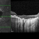
Optical coherence tomography with enhanced depth imaging of a 86-year-old male with optic nerve head drusen affecting his right eye. This patient has also been diagnosed with pseudoxanthoma elasticum and macular degeneration.
Photographer: Olivia Rainey
Imaging device: Heidelberg Spectralis
Condition/keywords: enhanced depth imaging, infrared image, macular degeneration, optic disc drusen, optic nerve, optical coherence tomography (OCT), pseudoxanthoma elasticum (PXE)
-
 SD OCT Comparison Analysis in Patient With (PXE)
Jun 10 2016 by Jordan Ball, DO
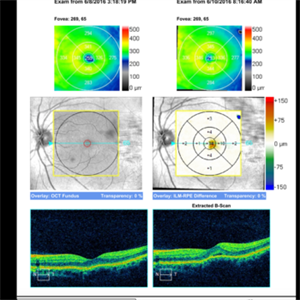
OCT scans taken two days apart. No subretinal fluid noted and patient asymptomatic on 6/8/2016 thus observation was suggested and elected. Patient called 6/9/16 complaining of new distortions and upon examination 6/10/16 new subretinal fluid was found. Patient subsequently treated with anti-vegf injection.
Condition/keywords: choroidal neovascularization (CNV), pseudoxanthoma elasticum (PXE)
-
 Fluorescein Angiography 1 in Patient With (PXE)
Jun 10 2016 by Jordan Ball, DO
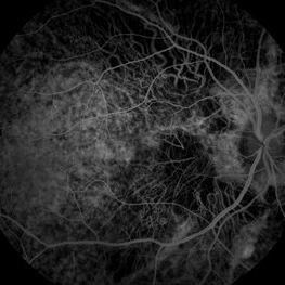
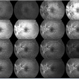
Fluorescein angiogram of patient with CNV from PXE showing angioid streaks and focal leakage at temporal edge of FAZ.
Condition/keywords: angioid streaks, choroidal neovascularization (CNV), pseudoxanthoma elasticum (PXE)
-
 Fluorescein Angiography 2 in Patient with (PXE)
Jun 10 2016 by Jordan Ball, DO
Late frames of fluorescein angiogram of patient with CNV from PXE showing Angioid streaks and focal leakage at temporal edge of FAZ.
Condition/keywords: angioid streaks, choroidal neovascularization (CNV), pseudoxanthoma elasticum (PXE)
-
 Angioid streaks from PXE
Jun 10 2016 by Jordan Ball, DO
Fundus photographs of left eye of patient with CNV associated to PXE showing angioid streaks.
Imaging device: Pseudoxanthoma Elasticum CNV
Condition/keywords: angioid streaks, pseudoxanthoma elasticum (PXE)
-
 Neck Skin of PXE Patient
Jun 10 2016 by Jordan Ball, DO
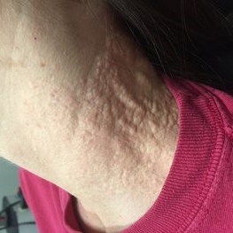
Neck skin of patient with pseudoxanthoma elasticum.
Condition/keywords: pseudoxanthoma elasticum (PXE)
-
 Antecubital Fossa Skin of PXE Patient
Jun 10 2016 by Jordan Ball, DO
Antecubital fossa skin of patient with pseudoxanthoma elasticum.
Condition/keywords: pseudoxanthoma elasticum (PXE)
-
 PXE
PXE
Dec 18 2014 by H. Michael Lambert, MD
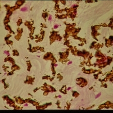
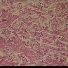
PXE - calcium stain.
Condition/keywords: pseudoxanthoma elasticum (PXE)
-
 PXE
PXE
Dec 18 2014 by H. Michael Lambert, MD
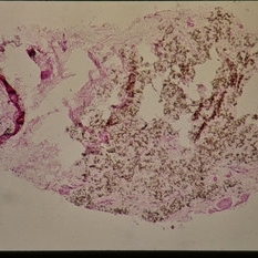
PXE - calcium stain.
Condition/keywords: pseudoxanthoma elasticum (PXE)
-
 PXE
PXE
-
 PXE
PXE
Dec 18 2014 by H. Michael Lambert, MD
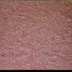
PXE - calcium stain.
Condition/keywords: pseudoxanthoma elasticum (PXE)
-
 PXE
PXE
-
 PXE
PXE
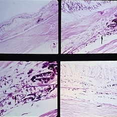
Dec 18 2014 by H. Michael Lambert, MD
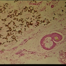
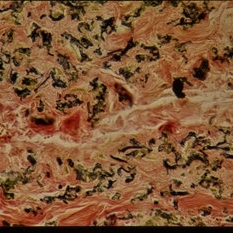
PXE - elastic stain.
Condition/keywords: pseudoxanthoma elasticum (PXE)
 Loading…