Search results (15 results)
-
 Herpetic Corneal Ulcer
Sep 24 2024 by DR Rohit Gupta
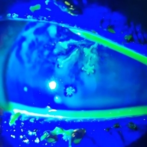
Slit lamp photograph of 32 year old male presented with herpetic corneal ulcer on staining with fluorescein dye under cobalt blue filted dendrits can be seen.
Photographer: Dr Rohit gupta
Imaging device: Samsung S21
Condition/keywords: corneal ulcer, dendritic keratitis, herpes dendrite, Herpes simplex infection, Herpes zoster, staining
-
 ARN (#3) This is comparison between the latest visit (left) and one week prior (which is the right photo, and same one as photo #2)
May 27 2019 by John S. King, MD
60-year-old African American female who had been treated for iridocyclitis for at least a week sent in for vitritis and a nasal fundus lesion. Complaints included redness, floaters, photophobia, and decreased vision. Husband had recent shingles. Acuity was 20/60-2 with IOP of 12, and small KP in Art's triangel, 1-2+ a/c cell, 2-3+ ant vit cell, diffuse arteriolar sheathing, multiple areas of retinal whitening in periphery and mid-periphery (see Photo #1). PCR of a/c was performed, and intravitreal GCV administered, and VACV 2g qid and ASA started.... PCR positive for HZV, pred taper was started two days after presentation as the infection had begun to stablize..... Five days from presentation the vision was 20/60, inflammation and areas of retinal whitening had improved (see Photo #2).... One week later acuity was 20/30, the a/c was quiet and KP resolved; ant vitreous cell decreased; and there was further improvement in retinal appearance without any signs of retinal holes or detachment; she is now on low dose maint VACV (see photo#3)
Photographer: Maysee Yang
Imaging device: Optos CA
Condition/keywords: acute retinal necrosis, Herpes zoster
-
 ARN (#2) Five Days Since Initial Visit
May 27 2019 by John S. King, MD
60-year-old African American female who had been treated for iridocyclitis for at least a week sent in for vitritis and a nasal fundus lesion. Complaints included redness, floaters, photophobia, and decreased vision. Husband had recent shingles. Acuity was 20/60-2 with IOP of 12, and small KP in Art's triangel, 1-2+ a/c cell, 2-3+ ant vit cell, diffuse arteriolar sheathing, multiple areas of retinal whitening in periphery and mid-periphery (see Photo #1). PCR of a/c was performed, and intravitreal GCV administered, and VACV 2g qid and ASA started.... PCR positive for HZV, pred taper was started two days after presentation as the infection had begun to stablize..... Five days from presentation the vision was 20/60, inflammation and areas of retinal whitening had improved (see Photo #2).... One week later acuity was 20/30, the a/c was quiet and KP resolved; ant vitreous cell decreased; and there was further improvement in retinal appearance without any signs of retinal holes or detachment; she is now on low dose maint VACV (see photo#3)
Photographer: Maysee Yang
Imaging device: Optos CA
Condition/keywords: acute retinal necrosis, Herpes zoster
-
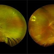
 ARN (#1) Initial Photo
May 27 2019 by John S. King, MD
60-year-old African American female who had been treated for iridocyclitis for at least a week sent in for vitritis and a nasal fundus lesion. Complaints included redness, floaters, photophobia, and decreased vision. Husband had recent shingles. Acuity was 20/60-2 with IOP of 12, and small KP in Art's triangel, 1-2+ a/c cell, 2-3+ ant vit cell, diffuse arteriolar sheathing, multiple areas of retinal whitening in periphery and mid-periphery (see Photo #1). PCR of a/c was performed, and intravitreal GCV administered, and VACV 2g qid and ASA started.... PCR positive for HZV, pred taper was started two days after presentation as the infection had begun to stablize..... Five days from presentation the vision was 20/60, inflammation and areas of retinal whitening had improved (see Photo #2).... One week later acuity was 20/30, the a/c was quiet and KP resolved; ant vitreous cell decreased; and there was further improvement in retinal appearance without any signs of retinal holes or detachment; she is now on low dose maint VACV (see photo#3)
Photographer: Maysee Yang
Imaging device: Optos CA
Condition/keywords: acute retinal necrosis, Herpes zoster
-
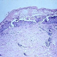 Slide 5-13
Feb 20 2019 by Lancaster Course in Ophthalmology
Intraepidermal bulla in herpes zoster of the skin.
Condition/keywords: intraepidermal bulla
-
 Slide 1-14
Feb 19 2019 by Lancaster Course in Ophthalmology
Russell body in center of field appears as a multilobed, pink body with faint nuclear remnants, while small lymphocytes and plasma cells are seen on the left, in chronic uveitis following herpes zoster. (H&E stain)
Condition/keywords: chronic uveitis, Herpes zoster, lymphocytes
-
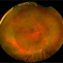 Acute Retinal Necrosis secondary to Herpes Zoster Ophthalmicus
Jan 9 2018 by Olivia Rainey
Ultra-wide field Optos pseudocolor montage of an 40-year-old female presenting with acute retinal necrosis secondary to herpes zoster ophthalmicus affecting her right eye.
Photographer: Olivia Rainey
Imaging device: Optos California
Condition/keywords: acute retinal necrosis, color fundus photograph, Herpes zoster, montage, Optos, ultra-wide field imaging
-
 Herpes Zoster
Jul 11 2013 by Jerald A. Bovino, MD
No history, vesicular skin rash along dermatome.
Condition/keywords: Herpes zoster, skin lesions
-
 Herpes Zoster
Jul 11 2013 by Jerald A. Bovino, MD
No history, vesicular skin rash along dermatome.
Condition/keywords: Herpes zoster, skin lesions
-
 Herpes Zoster
Jul 11 2013 by Jerald A. Bovino, MD
No history, vesicular skin rash along dermatome.
Condition/keywords: Herpes zoster, skin lesions
-
 Herpes Zoster
Jul 11 2013 by Jerald A. Bovino, MD
No history, vesicular skin rash along dermatome.
Condition/keywords: Herpes zoster, skin lesions
-
 Herpes Zoster
Jul 11 2013 by Jerald A. Bovino, MD
No history, vesicular skin rash along dermatome.
Condition/keywords: Herpes zoster, skin lesions
-
 Herpes Zoster
Jul 11 2013 by Jerald A. Bovino, MD
No history, vesicular skin rash along dermatome.
Condition/keywords: Herpes zoster, skin lesions
-
---thumb.jpg/image-square;max$300,300.ImageHandler) Brown/Mendis BJO 57:344, 1973
Brown/Mendis BJO 57:344, 1973
Feb 14 2013 by From the Collections of Thomas M. Aaberg, MD and Thomas M. Aaberg Jr., MD
reprints of figures 1 and 2 from the publication Brown and Mendis. Retinal arteritis complicating herpes zoster ophthalmicus. Br J Ophthalmol 1973;57:344-6. The left panel is a "fundus painting showing extensive exudate in areas of supply of narrowed and sheathed upper nasal and upper temporal retinal arterioles." The right panel is a fluorescein angiograph of the fundus, "demonstrating leakage of dye in area of exudation."
Condition/keywords: Herpes zoster, retinal arteriolar occlusion, retinal necrosis
-
 Herpes Zoster of Face
 Loading…