Search results (65 results)
-
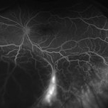
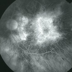
Central Serous Chorioretinopathy
Apr 15 2025 by Filip Kecer
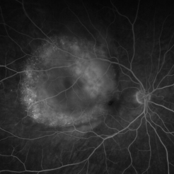
FA&ICG late phase of a young woman with CSCR
Photographer: Filip Kecer, Oftalmocentrum Betliarska, Bratislava, Slovakia
Imaging device: Spectralis, Heidelberg Engineering
Condition/keywords: central serous chorioretinopathy (CSCR), Central Serous Chorioretinopathy (CSR), FA late phase, indocyanine green (ICG) angiography
-
 Choroidal Hemangioma 4 Ways
Mar 13 2025 by Virginia Gebhart
Color fundus, FAF, late FA, late ICG of 64 year old male with choroidal hemangioma. Early hyperfluorescence with late leakage on FA, early hypercyanescence with late washout (25 min) on ICG.
Photographer: Virginia Gebhart, Retina Consultants of Carolina
Imaging device: Optos California
Condition/keywords: autofluorescence imaging, choroidal hemangioma, FA late phase, Fluorescein angiography, hemangioma, indocyanine green (ICG) angiography
-
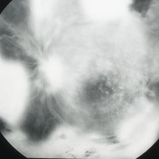
 Uveal Effusion Syndrome
Jan 7 2025 by Drew Mitchell
Optos FA Late of Uveal Effusion Syndrome
Photographer: Drew Mitchell, OCT-C
Imaging device: Optos California
Condition/keywords: FA late phase, Optos, uveal effusion
-
 Branch Retinal Vein Occlusion
Aug 22 2024 by Virginia Gebhart
Fluorescein angiogram of branch retinal vein occlusion in 75 year old female. Scattered microaneurysms with late CME and persistent SRF. Pt will consider laser treatment but is hesitant for injections at this time due to possible side effects.
Photographer: Virginia Gebhart
Imaging device: Optos California
Condition/keywords: branch retinal vein occlusion (BRVO), BRVO, cystoid macular edema (CME), FA, FA late phase, fluorescein angiogram (FA), macular edema, microaneurysms, retinal microaneurysms
-
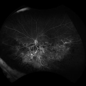 Ischemic HRVO with Macular Edema
Mar 7 2024 by Jenn Geelan
Optos FA of an 80 year old female.
Photographer: Jenn Geelan
Imaging device: Optos California
Condition/keywords: FA late phase, hemicentral retinal vein occlusion, ischemic CRVO, macular edema
-
 Acute Posterior Multifocal Placoid Pigment Epitheliopathy
Feb 20 2024 by Soobien Lee
Fluorescein angiogram of a 20-year-old caucasian female with viral prodrome and vision loss OS>OD secondary to Acute Posterior Multifocal Placoid Pigment Epitheliopathy (APPME). Early blockage with late hyperfluorescent leakage can be seen on fluorescein angiography of the left eye.
Photographer: Ashley Metzger, Elman Retina Group
Imaging device: Optos Ultra-Widefield Fluorescein Angiography
Condition/keywords: acute posterior multifocal placoid pigment epitheliopathy (APMPPE), bacilliary layer detachment, FA, FA late phase, FA late phase leakage, fluorescein angiogram (FA), Optos, uveitis, white dot syndrome
-
 Choroidal Melanoma
Oct 27 2023 by Virginia Gebhart
76 year old male with suspicious pigmented choroidal lesion with new collar button growth. Blocking defect and vascularity noted on FA
Photographer: Virginia Gebhart
Condition/keywords: FA late phase, fluorescein angiogram (FA), Fluorescein angiography, melanoma
-
 Polypoidal Choroidal Vasculopathy
Jul 20 2023 by Gregg T. Kokame, MD, MMM, FASRS
64 Year Old Male, with Polypoidal Choroidal Vasculopathy. Pre-op and Post-op PDT/Vabysmo Injection
Photographer: Jaclyn Pisano
Imaging device: Heidelberg Spectralis
Condition/keywords: FA late phase, indocyanine green (ICG) angiography, OCT, PDT, polypoidal choroidal vasculopathy (PCV), subretinal, subretinal fluid
-
 Cystoid Macular Degeneration
Feb 1 2023 by Kachelle Brown
Fluorescein Angiogram of a 56 year old woman with bilateral Cystoid Macular Degeneration. Patient vision was 20/60 OU.
Photographer: Kachelle Brown OMA, Retina Specialist of Michigan
Condition/keywords: cystoid macular degeneration, cystoid macular edema (CME), FA late phase, fluorescein angiogram (FA)
-
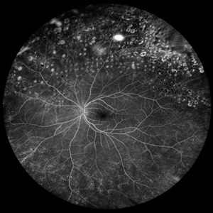
 Central Retinal Vein Occlusion with Severe Retinal Ischemia
Jan 19 2022 by Olivia Rainey
Ultra-widefield fluorescein angiogram of a 56-year-old male with a Central Retinal Vein Occlusion with Severe Retinal Ischemia affecting his right eye. The patient presented on 1/19/2022, sc20/20-2 vision in the right eye. The patient has had a good response to Eylea with complete resolution of edema. The physician is considering PRP to ischemic periphery in the future and given the degree of ischemia in both eyes, she recommends that the patient's PCP check carotid Doppler US.
Photographer: Olivia Rainey, OCT-C, COA
Imaging device: Optos California
Condition/keywords: central retinal vein occlusion (CRVO), FA late phase, fluorescein angiogram (FA), ischemic CRVO, Optos, retinal ischemia, ultra-wide field imaging
-
 Superotemporal Branch Retinal Vein Occlusion
Oct 3 2021 by Jesus Lozano, MD
Optos - Fluorescein Angiography widefield, ultra-high resolution angiography image of a 60 year-old man presented with blurred vision in the left eye. The patient was diagnosed with a superotemporal branch retinal vein occlusion.
Photographer: Yair Bet Yosef, Hadassah Medical Center. Israel
Imaging device: Optos
Condition/keywords: branch retinal vein occlusion (BRVO), fa, FA late phase leakage
-
 Breast cancer metastatic to choroid
Jul 13 2021 by Odette M. Houghton, MD
Late phase fluorescein angiogram of a 59-year-old female with a choroidal tumor secondary to metastatic breast cancer.
Photographer: David Saiz COT, Mayo Clinic Arizona
Imaging device: Optos California
Condition/keywords: breast cancer, FA late phase, metastatic cancer
-
 Syphilitic Uveitis
Apr 2 2020 by Olivia Rainey
Ultrawide-field fluorescein angiogram of a 42-year-old male with syphilitic uveitis affecting his right eye more than his left. Patient is HIV positive. He developed hearing loss and palm/leg/scalp rash prompting diagnosis of neurosyphilis, s/p IM and full IV course of 2.4 Mil PCN G, and finished this course 3/9/20. He admits to recent rectal bleeding with ongoing plan for colonoscopy 3/16/20. He has a history of extensive travel including London, Hong Kong, and Bangkok. His husband has also been treated with IV PCN G, however per chart review he has multiple sexual partners. Patient's vision was 20/20 in each eye.
Photographer: Olivia Rainey
Imaging device: Optos California
Condition/keywords: disc hyperfluorescence, FA late phase leakage, fluorescein angiogram (FA), fluorescein leakage, HIV, late phase, optic nerve edema, Optos, phelbitis, syphilis neuroretinopathy, ultra-wide field imaging, uveitis
-
 Coccidioides Choroiditis - Late FA
Mar 2 2020 by Scott C. Oliver, MD
Coccidioides choroiditis - late FA
Photographer: UCLA
Imaging device: Optos
Condition/keywords: choroiditis, coccidiomycosis, FA late phase
-
 Choroidal Detachment
Jan 6 2020 by Sarah Oelrich
Choroidal detachment
Photographer: Sarah Oelrich CRA COT OCT-C
Imaging device: Optos
Condition/keywords: choroidal detachment, detachment, FA late phase
-
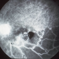
 Paracentral Acute Middle Maculopathy
Oct 22 2019 by Jeffrey G. Gross, MD, FASRS
FA of 75-year-old white male with 6 day history of acute vision loss. 20/40
Photographer: Tammy Mclaughlin
Imaging device: Zeiss Visucam
Condition/keywords: FA late phase, paracentral acute middle maculopathy, retinal ischemia
-
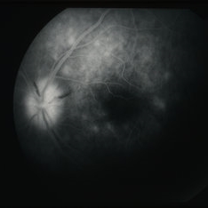
 Regional Choriocapillaris Atrophy
Jun 18 2019 by Gary R. Cook, MD, FACS
Late-phase (5 minutes) FA frame of the left eye of a 73-year-old white female with regional choriocapillaris atrophy showing light staining from intact choriocapillaris around the margins of the peripapillary and macular areas of RPE and choriocapillaris atrophy; V.A. = 20/100
Imaging device: Topcon VT-50
Condition/keywords: atrophy, choriocapillaris, FA late phase, fluorescein angiogram (FA), hereditary choroidal atrophy, hereditary choroidal dystrophy
-
 Uveitis with Laser FA
Apr 26 2019 by Carissa Hurdstrom
Uveitis with laser
Photographer: Carissa Hurdstrom
Imaging device: Optos
Condition/keywords: FA late phase, laser, uveitis
-
 Nonperfused BRVO with Collateral Vessels
Apr 8 2019 by Gary R. Cook, MD, FACS
Late-phase fluorescein angiogram image of the left eye of a 73-year-old African-American female with a nonperfused BRVO showing flow through the collateral vessels, marked loss of the capillary bed, disc leakage from some NVD, and ischemic staining of the retinal veins; V.A. = 20/70-1
Imaging device: Topcon VT-50
Condition/keywords: branch retinal vein occlusion (BRVO), capillary nonperfusion, collaterals, disc leakage, FA late phase, fluorescein angiogram (FA)
-
 Posterior Uveitis
Apr 8 2019 by Gary R. Cook, MD, FACS
Late-phase (219 seconds) fluorescein angiogram image of the left eye showing late staining of the optic disc and of numerous spots deep to the retina; also blocked fluorescence from the 2 NFL hemorrhages on the optic disc; V.A. = 20/20-1
Imaging device: Topcon VT-50
Condition/keywords: FA late phase, fluorescein angiogram (FA), posterior uveitis
-
 Syphilitic Chorioretinitis
Apr 8 2019 by Gary R. Cook, MD, FACS
Late-phase (10 minutes) fluorescein angiogram image of the posterior pole of the right eye of a 68 -year-old female patient with a diffuse, bilateral salt-and-pepper chorioretinitis secondary to syphilis; V.A. = 20/25
Imaging device: Topcon VT-50
Condition/keywords: chorioretinitis, FA late phase, fluorescein angiogram (FA), pseudo retinitis pigmentosa, syphilis
-
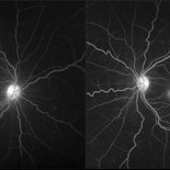
 X-Linked Retinitis Pigmentosa
Apr 2 2019 by Gary R. Cook, MD, FACS
Late-phase fluorescein angiogram image of the left eye of a 42-year old Vietnamese male with X-linked retinitis pigmentosa; V.A. = 20/25-3
Imaging device: Topcon VT-50
Condition/keywords: FA late phase, fluorescein angiogram (FA), retinitis pigmentosa, retinitis pigmentosa (RP) dystrophy
-
 X-Linked Retinitis Pigmentosa
Apr 2 2019 by Gary R. Cook, MD, FACS
Late-phase fluorescein angiogram image of the right eye of a 42-year old Vietnamese male with X-linked retinitis pigmentosa; V.A. = 20/25
Imaging device: Topcon VT-50
Condition/keywords: FA late phase, fluorescein leakage, retinitis pigmentosa, retinitis pigmentosa (RP) dystrophy
-
 Retinitis Pigmentosa with CME
Apr 2 2019 by Gary R. Cook, MD, FACS
Late-phase fluorescein angiogram frame from a 26-year-old white female with retinitis pigmentosa and CME OD; V.A. = 20/30
Imaging device: Topcon VT-50
Condition/keywords: cystoid macular edema (CME), FA late phase, fluorescein angiogram (FA), retinitis pigmentosa
-
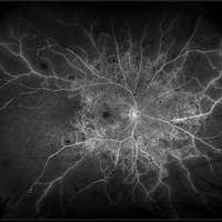
 Eales Disease
Apr 1 2019 by Gary R. Cook, MD, FACS
Late-phase fluorescein angiogram image of the left eye of a 23-year-old Vietnamese female with Eales Disease showing extensive dye leakage from multiple areas of NVE and from some NVD.
Imaging device: Topcon VT-50
Condition/keywords: Eales disease, FA late phase, fluorescein angiogram (FA), neovascularization elsewhere (NVE)
 Loading…