-
 Macular Pucker
Jan 7 2020 by RAFAEL REIS PEREIRA, MD
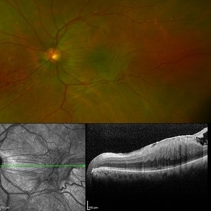
A clinical grading system was proposed by Gass in 1987 describe the different stages of the epiretinal membrane. Grade 2 Macular pucker consists of a thick fibroglial membrane that contracts and produces obscuration of underlying vessels and marked full-thickness retinal distortion. Sometimes associated with cotton-wool spots, exudates, blot hemorrhages, microaneurysms, and cystoid macular edema.
Photographer: Rafael Reis, Retina Clinic - Brazil
Condition/keywords: macular pucker
-
 Retinal Angiomatous Proliferation RAP
Mar 11 2020 by RAFAEL REIS PEREIRA, MD
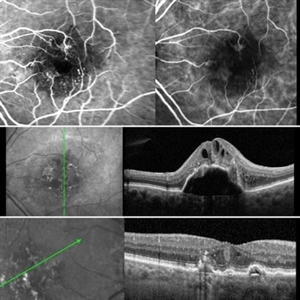
Retinal angiomatous proliferation (RAP) is a unique variant of neovascular age-related macular degeneration. Published studies have estimated that up to 15% of patients with neovascular age-related macular degeneration have RAP. Clinical features frequently associated with RAP include bilateral disease, presence of pigment epithelial detachments, and reticular pseudodrusen. RAP is more frequently associated with the development of retinal pigment epithelial tears and geographic atrophy that can lead to severe vision loss. We present a stereo fluorescein angiography and ICG (upper right and left image respectively) and OCT of left and right eye (middle and inferior image) of a RAP choroidal neovascularization in an 89-year-old patient.
Photographer: Rafael Reis Pereira
Imaging device: HRA Heildelberg Spectralis
Condition/keywords: retinal angiomatous proliferation (RAP)
-
 Valsalva Retinopathy
Feb 23 2021 by RAFAEL REIS PEREIRA, MD
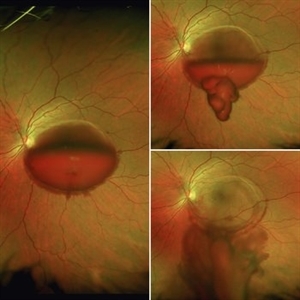
Valsalva retinopathy is a specific form of retinopathy characterized by pre-retinal hemorrhages secondary to raised intrathoracic pressure. This is a 31-year-old female who had breast implant surgery and complained of low VA in her left eye since the procedure. The patient had a large subhyaloid hemorrhage and we performed Nd YAG laser restoring 20/20 vision in the 4th-day post-treatment.
Condition/keywords: retina, valsalva retinopathy
-
 Acute Syphilitic Posterior Placoid Chorioretinitis
May 4 2021 by RAFAEL REIS PEREIRA, MD
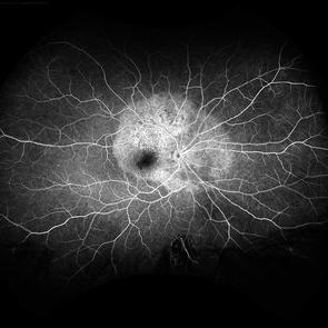
A 31-year-old patient with a complaint of photophobia and low visual acuity OD in the previous three weeks. BCVA was 20/60 and 20/20 The fundus examination revealed a placoid white lesion in the posterior pole and vitreous cells in the right eye. The left eye was unremarkable. Fluorescein angiography reveals hyperfluorescent plaque with distinctive “leopard spots” hypofluorescence.
Imaging device: Opto California
Condition/keywords: acute syphilitic posterior placoid chorioretinitis
A project from the American Society of Retina Specialists