-
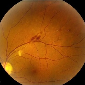
 Arterial Occlusion
Jul 14 2013 by Jason S. Calhoun
Patient in with vision loss in the lower quadrant of his visual field. Fundus photo shows arterial occlusion superior to the optic nerve
Photographer: Jason S. Calhoun, Department of Ophthalmology, Mayo Clinic Jacksonville, Florida
Imaging device: TOPCON TRC 50-EX
Condition/keywords: branch retinal artery occlusion (BRAO)
-
---thumb.JPG/image-square;max$300,300.ImageHandler) Central Retinal Vein Occlusion 1
Central Retinal Vein Occlusion 1
Jul 14 2013 by Jason S. Calhoun
Central Retinal Vein Occlusion.
Photographer: Jason S. Calhoun, Department of Ophthalmology, Mayo Clinic Jacksonville, Florida
Imaging device: TOPCON TRC 50-EX
Condition/keywords: central retinal vein occlusion (CRVO)
-
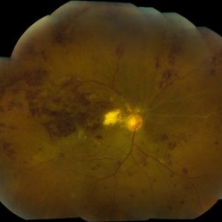
 Central Retinal Vein Occlusion
Jul 14 2013 by Jason S. Calhoun
Central retinal vein occlusion.
Photographer: Jason S. Calhoun, Department of Ophthalmology, Mayo Clinic Jacksonville, Florida
Imaging device: TOPCON TRC 50-EX
Condition/keywords: central retinal vein occlusion (CRVO)
-
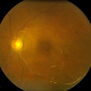
 Branch Artery Occlusion
Jul 14 2013 by Jason S. Calhoun
Superior branch artery occlusion in the left eye
Photographer: Jason S. Calhoun, Department of Ophthalmology, Mayo Clinic Jacksonville, Florida
Imaging device: TOPCON TRC 50-EX
Condition/keywords: branch retinal artery occlusion (BRAO)
-
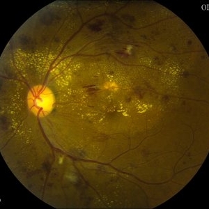
 Calcium Deposits of Central Retinal Vein
Jul 14 2013 by Jason S. Calhoun
Vein occlusion with severe blockage inferiorly. Calcium deposits seen through the veins branching out with thinning of the retina.
Photographer: Jason S. Calhoun, Department of Ophthalmology, Mayo Clinic Jacksonville, Florida
Imaging device: TOPCON TRC 50-EX
Condition/keywords: branch retinal vein occlusion (BRVO)
-
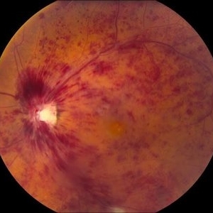
 Combined Artery and Vein Occlusion
Jun 27 2013 by Jason S. Calhoun
Patient comes in with decreased vision in both eyes. VA 20/200-OD, 20/60-OS. Fundus exam shows great amount of macular edema due to artery and vein occlusions. There is some neovascularization on the optic nerve in the right eye. Patient was treated with Eylea injection in the left eye today and will return for Eylea injection in the right eye.
Photographer: Jason S. Calhoun, Mayo Clinic Jacksonville, Florida
Imaging device: TOPCON TRC 50-EX
Condition/keywords: branch retinal artery occlusion (BRAO), branch retinal vein occlusion (BRVO)
-
 Combined Artery and Vein Occlusion
Jun 27 2013 by Jason S. Calhoun
Patient came in with decreased vision in both eyes. VA 20/200-OD, 20/60-OS. Fundus exam shows great amount of macular edema due to artery and vein occlusions. There is some neovascularization on the optic nerve in the right eye. Patient was treated with Eylea injection in the left eye today and will return for Eylea injection in the right eye.
Photographer: Jason S. Calhoun, Mayo Clinic Jacksonville, Florida
Imaging device: TOPCON TRC 50-EX
Condition/keywords: branch retinal vein occlusion (BRVO), central retinal vein occlusion (CRVO)
A project from the American Society of Retina Specialists