-
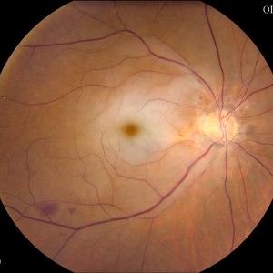
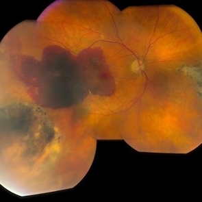 Retinal Hemorrhage
Jun 29 2013 by Jason S. Calhoun
Patient has history of macular degeneration (wet). Also has had vitrectomy in the left eye for a retinal detachment. Chief complaint was a spot in vision, right eye. Patient's VA was hand motion in the right eye. Retinal hemorrhage was present. Avastin injection was carried out and patient will have a follow up in one month.
Photographer: Jason S. Calhoun, Mayo Clinic Jacksonville, Florida
Imaging device: TOPCON TRC 50-EX
Condition/keywords: retinal hemorrhage
-
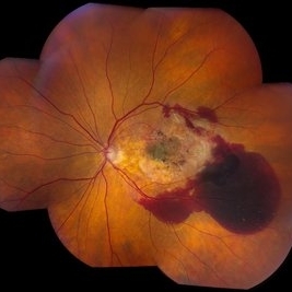 Macular Degeneration With Hemorrhage
Jun 29 2013 by Jason S. Calhoun
Patient comes in for follow up exam for wet AMD. Fundus photography reveals retinal hemorrhage temporally to the macula. Patient was not treated. Wait to see if the blood will absorb on its own.
Photographer: Jason S. Calhoun, Mayo Clinic Jacksonville, Florida
Imaging device: TOPCON TRC 50-EX
Condition/keywords: choroidal neovascularization (CNV)
-
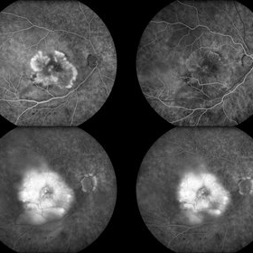 Wet Age-Related Macular Degeneration 4-Up
Jun 29 2013 by Jason S. Calhoun
Patient complains of gray spot in vision in the right eye. Fluorescein angiogram shows abnormal blood vessels leaking. Patient was treated with eylea Injection and will return for follow up in 1 month.
Photographer: Jason S. Calhoun, Mayo Clinic Jacksonville, Florida
Imaging device: TOPCON TRC 50-EX
Condition/keywords: age-related macular degeneration (AMD), choroidal neovascularization (CNV)
-
---thumb.JPG/image-square;max$300,300.ImageHandler) FAF of Macular Degeneration
FAF of Macular Degeneration
Jul 12 2013 by Jason S. Calhoun
Autofluresence or FAF photo of bilateral age related macular degeneration in both eyes.
Photographer: Jason S. Calhoun, Department of Ophthalmology, Mayo Clinic Jacksonville, Florida
Condition/keywords: autofluorescence imaging
-
---thumb.JPG/image-square;max$300,300.ImageHandler) FAF of Macular Degeneration
FAF of Macular Degeneration
Jul 12 2013 by Jason S. Calhoun
Autofluorescence or FAF photo of bilateral age related macular degeneration in both eyes.
Photographer: Jason S. Calhoun, Department of Ophthalmology, Mayo Clinic Jacksonville, Florida
Condition/keywords: autofluorescence imaging
-
---thumb.JPG/image-square;max$300,300.ImageHandler) Retinal Hemorrhage
Retinal Hemorrhage
Jul 13 2013 by Jason S. Calhoun
Retinal hemorrhage due to macular degeneration.
Photographer: Jason S. Calhoun, Department of Ophthalmology, Mayo Clinic Jacksonville, Florida
Condition/keywords: retinal hemorrhage
-
---thumb.JPG/image-square;max$300,300.ImageHandler) Disciform Scar
Disciform Scar
Jul 13 2013 by Jason S. Calhoun
Poor central vision in the left eye due to macular degeneration. Disciform scar.
Photographer: Jason S. Calhoun, Department of Ophthalmology, Mayo Clinic Jacksonville, Florida
Imaging device: TOPCON TRC 50-EX
Condition/keywords: disciform scar, macular degeneration
-
---thumb.JPG/image-square;max$300,300.ImageHandler) "Flower" Macular Degeneration (Wet)
"Flower" Macular Degeneration (Wet)
Jul 13 2013 by Jason S. Calhoun
Patient with (wet) macular degeneration in the left eye. Notice the "flower" shape abnormal blood vessels staining.
Photographer: Jason S. Calhoun, Department of Ophthalmology, Mayo Clinic Jacksonville, Florida
Imaging device: TOPCON TRC 50-EX
Condition/keywords: choroidal neovascularization (CNV)
-
---thumb.JPG/image-square;max$300,300.ImageHandler) Choroidal Neovascularization (CNV)
Choroidal Neovascularization (CNV)
Jul 13 2013 by Jason S. Calhoun
Fluorescein Angiography shows late staining which represents classic CNV.
Photographer: Jason S. Calhoun, Department of Ophthalmology, Mayo Clinic Jacksonville, Florida
Imaging device: TOPCON TRC 50-EX
Condition/keywords: choroidal neovascularization (CNV)
-
---thumb.JPG/image-square;max$300,300.ImageHandler) Choroidal Neovascularization (CNV)
Choroidal Neovascularization (CNV)
Jul 13 2013 by Jason S. Calhoun
Fluorescein angiography shows early staining which represents classic CNV.
Photographer: Jason S. Calhoun, Department of Ophthalmology, Mayo Clinic Jacksonville, Florida
Imaging device: TOPCON TRC 50-EX
Condition/keywords: choroidal neovascularization (CNV)
-
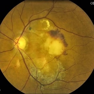
---thumb.JPG/image-square;max$300,300.ImageHandler) Dry Macular Degeneration With Hemorrhage
Dry Macular Degeneration With Hemorrhage
Jul 13 2013 by Jason S. Calhoun
Pigment changes in the macula with hemorrhages present temporally.
Photographer: Jason S. Calhoun, Department of Ophthalmology, Mayo Clinic Jacksonville, Florida
Imaging device: TOPCON TRC 50-EX
Condition/keywords: dry age-related macular degeneration (dry AMD)
-
 Choroidal Neovascularization (CNV)
Jul 13 2013 by Jason S. Calhoun
Active CNV in middle aged black female. Proceeded with Avastin injection.
Photographer: Jason S. Calhoun, Department of Ophthalmology, Mayo Clinic Jacksonville, Florida
Imaging device: TOPCON TRC 50-EX
Condition/keywords: choroidal neovascularization (CNV)
-
 CRAO With Embolis
Jul 16 2013 by Jason S. Calhoun
Patient with active leaking of AMD in the left eye. VA is 20/40 in the left eye. Patient was treated with Eylea and will return in 1-month for follow up.
Photographer: Jason S. Calhoun, Department of Ophthalmology, Mayo Clinic Jacksonville, Florida
Imaging device: TOPCON TRC 50-EX
-
---thumb.JPG/image-square;max$300,300.ImageHandler) Macular Degeneration With Hemorrhage 1
Macular Degeneration With Hemorrhage 1
Jul 16 2013 by Jason S. Calhoun
Patient with active leaking of AMD in the left eye. VA is 20/40 in the left eye. Patient was treated with Eylea and will return in 1-month for follow up.
Photographer: Jason S. Calhoun, Department of Ophthalmology, Mayo Clinic Jacksonville, Florida
Imaging device: TOPCON TRC 50-EX
-
---thumb.JPG/image-square;max$300,300.ImageHandler) Macular Degeneration With Hemorrhage
Macular Degeneration With Hemorrhage
Jul 16 2013 by Jason S. Calhoun
Patient with active leaking of AMD in the left eye. VA is 20/40 in the left eye. Patient was treated with Eylea and will return in 1-month for follow up.
Photographer: Jason S. Calhoun, Department of Ophthalmology, Mayo Clinic Jacksonville, Florida
Imaging device: TOPCON TRC 50-EX
A project from the American Society of Retina Specialists