-
 Central Retinal Vein Occlusion with Foveal Hemorrhage
Apr 17 2025 by Malvika Singh
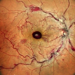
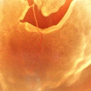
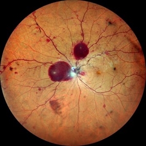
Fundus photograph of a 41 year-old, male, with a central retinal vein occlusion and a foveal sub-internal limiting membrane hemorrhage.
Photographer: Dr Malvika Singh, Retina Foundation, Ahmedabad, India
Imaging device: Mirante SLO/OCT
Condition/keywords: central retinal vein occlusion (CRVO), macular hemorrhage
-
 Sub-Internal Limiting Membrane Hemorrhage
Apr 17 2025 by Malvika Singh
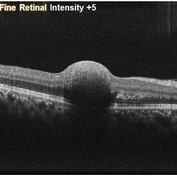
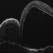
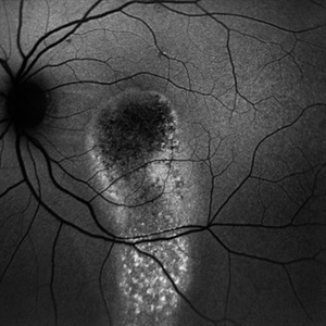
OCT of a 41 year-old, male, with a central retinal vein occlusion and a foveal sub-internal limiting membrane hemorrhage.
Photographer: Dr Malvika Singh, Retina Foundation, Ahmedabad, India
Imaging device: Mirante SLO/OCT
Condition/keywords: optical coherence tomography (OCT), SUB ILM hemorrhage
-
 Retinitis Pigmentosa with Macular Hole with Posterior Subcapsular Cataract
Apr 28 2025 by Malvika Singh
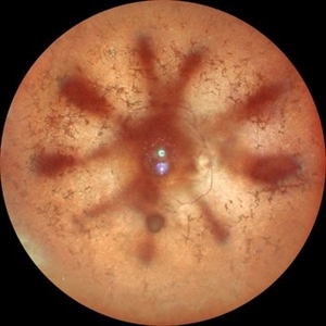
Fundus photograph of the right eye of a 31 year old with retinitis pigmentosa with a macular hole, showing the shadow of posterior subcapsular cataract over the fundus.
Photographer: Dr Malvika Singh, Retina Foundation, Ahmedabad, India
Imaging device: Mirante SLO/OCT
Condition/keywords: macular hole, posterior subcapsular cataract, retinitis pigmentosa
-
 Retinitis Pigmentosa with Macular Hole with Posterior Subcapsular Cataract
Apr 28 2025 by Malvika Singh
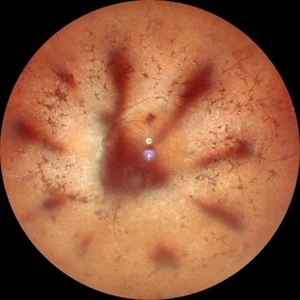
Fundus photograph of the left eye of a 31 year old with retinitis pigmentosa, showing the shadow of posterior subcapsular cataract over the fundus.
Photographer: Dr Malvika Singh, Retina Foundation, Ahmedabad, India
Imaging device: Mirante SLO/OCT
Condition/keywords: posterior subcapsular cataract, retinitis pigmentosa
-
 A Vessel That Would Not Let Go
May 5 2025 by Malvika Singh
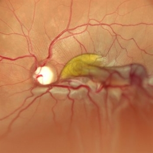
Fundus photograph of a retinal detachment showing a horse shoe shaped tear and a bridging vessel.
Photographer: Dr Tejaswita Verma, Retina Foundation, Ahmedabad, India
Imaging device: Mirante SLO/OCT
Condition/keywords: bridging vessel, horseshoe tear
-
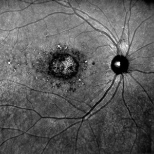
 Circinate Mark at the Macula — a Lasting Trace of Branch Retinal Vein Occlusion
May 13 2025 by Malvika Singh
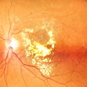
Fundus photograph of a 60 year old with a sclerosed vessel with hard exudates at the macula showing circinate retinopathy after an episode of branch retinal vein occlusion.
Photographer: Dr Malvika Singh, Retina Foundation, Ahmedabad, India
Imaging device: Mirante SLO/OCT
Condition/keywords: branch retinal vein occlusion (BRVO), circinate retinopathy
-
 Vogt Kayanagi Harada Disease
May 26 2025 by Malvika Singh
OCT image of the retina showing SRF in case of an exudative retinal detachment and bacillary layer detachment
Photographer: Dr Malvika Singh, Retina Foundation, Ahmedabad, India
Imaging device: Mirante SLO/OCT
Condition/keywords: OCT, Vogt-Koyanagi-Harada
-
Central Retinal Artery Pulsations
May 27 2025 by Malvika Singh
Fundus video showing pulsations of the central retinal artery at the excavated optic nerve head.
Condition/keywords: Central retinal artery, Excavated Disc, Fundus Video
-
 Traction in Proliferative Diabetic Retinopathy
Jun 9 2025 by Malvika Singh
Fundus photograph of a 44 year old with uncontrolled diabetes showing fibrovascular proliferation and traction with details of disc and macula obscured with sclerosed vessels in the periphery.
Photographer: Dr Malvika Singh, Retina Foundation, Ahmedabad, India
Imaging device: Mirante SLO/OCT
Condition/keywords: proliferative diabetic retinopathy (PDR), TRACTION
-
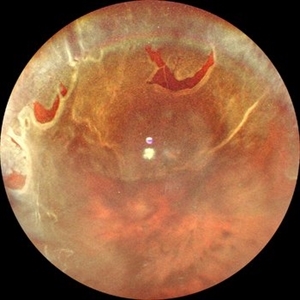 Retinal Detachment with Multiple Horse Shoe Shaped Tears
Jul 14 2025 by Malvika Singh
Fundus photograph of a 46 year old showing a retinal detachment with multiple peripheral horse show shaped tears.
Photographer: Dr Malvika Singh, Retina Foundation, Ahmedabad, India
Imaging device: Mirante SLO/OCT
Condition/keywords: horseshoe tear, retinal detachment
-
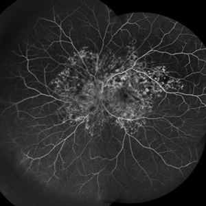
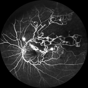 Branch Retinal Vein Occlusion
Jul 23 2025 by Malvika Singh
Fluorescein angiogram of a 52 year old man showing capillary non perfusion areas and leakages along the superotemporal arcade and at the macula.
Photographer: Dr Malvika Singh, Retina Foundation, Ahmedabad, India
Imaging device: Mirante SLO/OCT
Condition/keywords: branch retinal vein occlusion (BRVO), CNP areas, macular edema
-
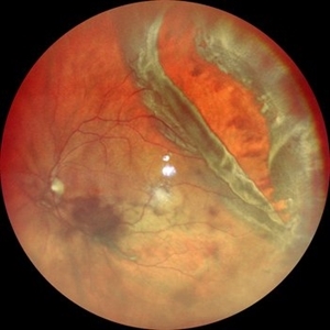 Jaws of Detachment
Jul 25 2025 by Malvika Singh
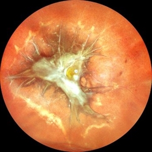
Fundus photograph of a 50 year old with a giant retinal tear and vitreous hemorrhage.
Photographer: Dr Malvika Singh, Retina Foundation, Ahmedabad, India
Imaging device: Mirante SLO/OCT
Condition/keywords: giant retinal tear
-
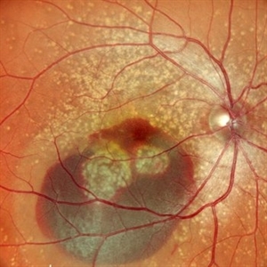 A Crack in the Honeycomb
Jul 28 2025 by Malvika Singh
Fundus photograph of a 44 year old with Doyne's Honeycomb Retinal Dystrophy (autosomal dominant drusen) and a subretinal bleed.
Photographer: Dr Malvika Singh, Retina Foundation, Ahmedabad, India
Imaging device: Mirante SLO/OCT
Condition/keywords: Doyne's Honeycomb, retinal dystrophy, subretinal blood
-
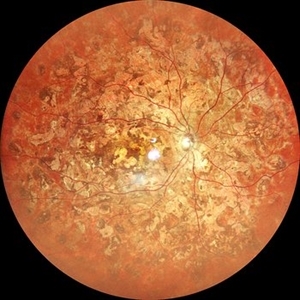 Ghost Map Retina
Aug 4 2025 by Malvika Singh
Fundus photograph of a 50 year old male showing extensive chorioretinal scarring.
Photographer: Dr Malvika Singh, Retina Foundation, Ahmedabad, India
Imaging device: Mirante SLO/OCT
Condition/keywords: healed choroiditis
-
 Stars of Stargardt
Aug 4 2025 by Malvika Singh
Infrared fundus photograph of a 22 year old female with Stargardt's disease.
Photographer: Dr Malvika Singh, Retina Foundation, Ahmedabad, India
Imaging device: Mirante SLO/OCT
Condition/keywords: infrared image, Stargardt disease
-
 Snaking Away
Sep 1 2025 by Malvika Singh
Fluorescein angiography montage of a 45 year old man showing areas of staining in a case of healed choroiditis.
Photographer: Dr Malvika Singh, Retina Foundation, Ahmedabad, India
Imaging device: Mirante SLO/OCT
Condition/keywords: healed choroiditis, serpiginous choroiditis
-
 Strained Retina
Sep 27 2025 by Malvika Singh
Fundus photograph of a 44 year old male showing hemorrhages at different layers.
Photographer: Dr Malvika Singh, Retina Foundation, Ahmedabad, India
Imaging device: Mirante SLO/OCT
Condition/keywords: valsalva retinopathy
-
 Strained Retina
Sep 27 2025 by Malvika Singh
Fundus photograph of a 44 year old male showing hemorrhages at different layers.
Photographer: Dr Malvika Singh, Retina Foundation, Ahmedabad, India
Imaging device: Mirante SLO/OCT
Condition/keywords: valsalva retinopathy
-
 Serous River of Light
Sep 30 2025 by Malvika Singh
Fundus autofluorescence of a 55 year old man with history of chronic central serous chorioretinopathy showing an old gravitational track.
Photographer: Dr Malvika Singh, Retina Foundation, Ahmedabad, India
Imaging device: Mirante SLO/OCT
Condition/keywords: central serous retinopathy (CSR)
-
 Folded Macula
Oct 13 2025 by Malvika Singh
Fundus photograph of a 25 year old male showing retinal detachment with macula off with retinal folds at the macula.
Photographer: Dr Malvika Singh, Retina Foundation, Ahmedabad, India
Imaging device: Mirante SLO/OCT
Condition/keywords: retinal detachment
-
 Multilayer Trauma
Nov 3 2025 by Malvika Singh
Fundus photograph of a 34 year old following trauma showing a choroidal rupture, a sub RPE and sub retinal bleed.
Photographer: Dr Malvika Singh, Retina Foundation, Ahmedabad, India
Imaging device: Mirante SLO/OCT
Condition/keywords: Choroidal Rupture, subretinal hemorrhage
-
 Optic Disc Drusen in Rod Cone Dystrophy
Nov 3 2025 by Malvika Singh
Fundus autofluorescence of a 22 year old male with rod cone dystrophy with hyperautofluorescent disc drusen.
Photographer: Dr Malvika Singh, Retina Foundation, Ahmedabad, India
Imaging device: Mirante SLO/OCT
Condition/keywords: optic disc drusen, Rod cone dystrophy
-
 Filigree Networks
Nov 14 2025 by Malvika Singh
Fundus photograph of a 31 year old male with type 1 diabetes mellitus showing neovascularisation along the superotemporal arcade.
Photographer: Dr Malvika Singh, Retina Foundation, Ahmedabad, India
Imaging device: Mirante SLO/OCT
Condition/keywords: neovascularization (NV), proliferative diabetic retinopathy (PDR)
-
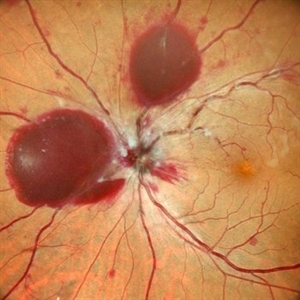
 The Bonus Vessel's Betrayal
Dec 8 2025 by Malvika Singh
Fundus photograph of a healthy 46 year old male with cilioretinal artery occlusion, macular ischemia and cherry red spot with normal surrounding retina.
Photographer: Malvika Singh
Imaging device: Mirante SLO Imaging Device
Condition/keywords: cilioretinal artery occlusion
-
 Layered Echoes of a Lost Lens
Dec 16 2025 by Malvika Singh
B scan of a 31 year old male, showing a dislocated crystalline lens in the vitreous.
Photographer: Malvika Singh
Condition/keywords: dislocated crystalline lens
-
Unanchored
Dec 16 2025 by Malvika Singh
Dynamic B-scan showing free movements of the dislocated lens in the vitreous cavity post trauma.
Condition/keywords: dislocated lens
-
The B-scan Ballet
Dec 16 2025 by Malvika Singh
Dynamic B-scan showing post-traumatic dislocated lens in the vitreous, freely moving in the cavity with eye movements.
Condition/keywords: dislocated lens
A project from the American Society of Retina Specialists