-
 Cannula Tip Pressure
Mar 25 2025 by Robert Andrew Sisk, MD, FACS, FASRS
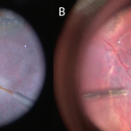
Color stills from surgical videos of subretinal delivery of gene augmentation therapy with A) voretigene neparvovec-ryzl and B) laru-zova. In the left panel, the cannula is slightly bent, and the retina and RPE are blanched white around the cannula tip engagement. The bleb was challenging to form in this patient with advanced retinal degeneration, and the bleb is shallow and mostly clear. In the right panel, the cannula tip is gently engaged, the cannula is straight, and it follows the retinotomy as the retina is elevated by the injection fluid.
Imaging device: Leica Proveo 8
Condition/keywords: gene therapy, genetic disorder, Leber's congenital amaurosis, retinitis pigmentosa, subretinal injection
-
 Completed Bleb with OCT Through Fovea
Mar 25 2025 by Robert Andrew Sisk, MD, FACS, FASRS
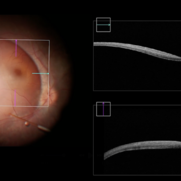
Color still from surgical video of subretinal delivery of laru-zova for X-linked retinitis pigmentosa. Live optical coherence tomography (OCT) with foveal tracking via the embedded software in the operating microscope allows monitoring foveal integrity for signs of stress. The contour of the fovea does not exceed the curvature of the bleb (e.g. no inversion). The tangential cannula angle facilitated steering of the bleb posteriorly. The bleb covers essentially the entire macula, which is the target area.
Imaging device: Zeiss Artevo 800
Condition/keywords: gene therapy, genetic disorder, optical coherence tomography (OCT), retinitis pigmentosa, subretinal injection
-
 Bleb Migration With FAX
Mar 25 2025 by Robert Andrew Sisk, MD, FACS, FASRS
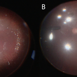
Color stills from surgical video after subretinal delivery of gene augmentation therapy with voretigene neparvovec-rzyl A) before and B) after fluid-air exchange (FAX). The blebs were between 0.5- and 1-disc diameters from the fovea. After FAX, they gradually extended beneath the fovea and eventually merged. This spared the fovea the trauma from the injection pressure of subretinal injection while allowing treatment to the area.
Imaging device: Leica Proveo 8
Condition/keywords: Fluid-Air Exchange, Gene Therapy, genetic disorder, genetics, Subretinal Injection
A project from the American Society of Retina Specialists