-
 Vortex-pattern Exudative Retinal Detachment
Feb 22 2025 by CUI YUELING
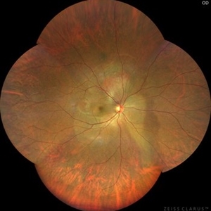
Patient: Male, 40 years old. Chief Complaint: Blurred vision and metamorphopsia in the left eye for more than 10 days. Past Medical History Hypertension for 4 years, with a highest recorded blood pressure of 160/80 mmHg. Currently controlled with oral "Nifedipine Sustained-Release Tablets, 2 tablets daily." Long-term history of heavy alcohol consumption and smoking. Ophthalmic Examination: Visual Acuity: Right eye (OD): 0.4 (uncorrected, no improvement with correction). Left eye (OS): 0.5 (-1.5DS = 1.0). Intraocular Pressure (IOP): OD: 15 mmHg. OS: 17 mmHg. Anterior Segment:Unremarkable. Fundus Examination: Right eye: Optic disc margins are clear, with a slightly reddish hue. Cup-to-disc ratio (C/D) = 0.2. A scalloped, orange-red elevated lesion is observed superior to the optic disc, with anterior displacement of the focal point. This is accompanied by a secondary, turbine-like exudative retinal detachment centered around the optic disc, involving the macula. The macular region shows scattered punctate yellow-white exudates. Diagnosis: Choroidal hemangioma with secondary exudative retinal detachment(OD).
Photographer: Cui yueling The First People's Hospital of Zunyi, Guizhou, Zunyi, China
Imaging device: Zeiss Clarus 500
Condition/keywords: choroidal hemangioma, exudative retinal detachment
-
 Commotio Retinae
Jun 10 2025 by CUI YUELING
The patient presented 2 hours after sustaining a left eye injury caused by a stick. Visual acuity in the left eye was 0.2 without improvement upon correction, and intraocular pressure measured 15 mmHg. Examination of the anterior segment revealed ciliary conjunctival injection accompanied by patchy subconjunctival hemorrhage. The corneal surface remained smooth, and the anterior chamber was deep with hyphema characterized by blood-tinged aqueous humor predominantly settled inferiorly. The pupil was slightly irregular, approximately 3 mm in diameter, with a superotemporal notch; pupillary light reflex was intact. The lens appeared clear. Fundus examination showed well-defined optic disc margins with normal coloration and a cup-to-disc ratio of 0.2. Retinal arteries and veins were normally distributed with an artery-to-vein ratio of 2:3. At the posterior pole, the foveal reflex exhibited concentric ripple-like changes centered on the fovea, accompanied by localized pigment attenuation and reduced reflex intensity. Irregular reflectivity was noted in the superotemporal and inferotemporal nerve fiber layers.
Photographer: Yueling Cui
Imaging device: Zeiss Clarus 500
Condition/keywords: commotio retinae
-
 Congenital Retinal Macrovessel
Jan 9 2026 by CUI YUELING
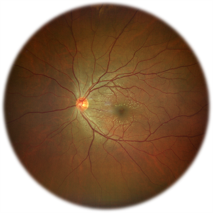
A 34-year-old female underwent fundus screening at an endocrinology clinic. She had a 5-year history of type 2 diabetes mellitus. Best-corrected visual acuity (BCVA) was 1.0 in both eyes. Anterior segment examination was unremarkable. Fundus examination showed mild nasal tilting of the optic disc in the right eye. The retinal artery-to-vein ratio was approximately 2:3. Notably, a superotemporal branch retinal artery was observed crossing the horizontal raphe and obliquely traversing the perifoveal capillary arcade within the foveal avascular zone (FAZ).
Photographer: Cui yueling The First People's Hospital of Zunyi, Guizhou, Zunyi, China
Imaging device: Zeiss Clarus 500
Condition/keywords: Congenital Retinal Macrovessel, OCT
A project from the American Society of Retina Specialists