-
 Leukemic Retinopathy
Nov 27 2024 by Ramses Rosales-Diaz
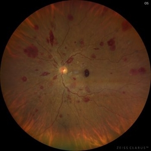
Fundus photograph of a 48-year-old woman with venous dilatation and tortuosity, flame-shaped and intraretinal hemorrhages, Roth spots and sub-ILM hemorrhage. Her complete blood count reports 425,540 lymphocytes/microliter, and the blood smear reveals Gumprecht shadows and numerous lymphocytes with nuclei exhibiting hypercondensed chromatin. She is diagnosed with chronic lymphocytic leukemia and receives appropriate treatment from the hematology team
Photographer: Ramses Rosales-Diaz, Asociación Para Evitar la Ceguera en México
Imaging device: Clarus 700
Condition/keywords: leukemia, sub ILM hemorrhage, white centered retinal hemorrhage (Roth Spot)
-
 Leukemic Retinopathy
Nov 27 2024 by Ramses Rosales-Diaz
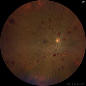
Fundus photograph of a 48-year-old woman showing venous dilatation and tortuosity, flame-shaped hemorrhages, intraretinal hemorrhages, sub-ILM hemorrhages, and Roth spots. Her complete blood count shows 425,540 lymphocytes/microliter, and the blood smear reveals Gumprecht shadows along with numerous lymphocytes with hypercondensed chromatin in their nuclei. She is diagnosed with chronic lymphocytic leukemia and receives appropriate treatment from the hematology team.
Photographer: Ramses Rosales-Diaz, Asociación Para Evitar la Ceguera en México
Imaging device: Zeiss Clarus 700
Condition/keywords: leukemia, sub ILM hemorrhage, white centered retinal hemorrhage (Roth Spot)
-
 Leukemic Retinopathy
Nov 27 2024 by Ramses Rosales-Diaz
Fundus photograph of a 48-year-old woman showing venous dilatation and tortuosity, flame-shaped hemorrhages, intraretinal hemorrhages, sub-ILM hemorrhages, and Roth spots. Her complete blood count shows 425,540 lymphocytes/microliter, and the blood smear reveals Gumprecht shadows along with numerous lymphocytes with hypercondensed chromatin in their nuclei. She is diagnosed with chronic lymphocytic leukemia and receives appropriate treatment from the hematology team.
Photographer: Ramses Rosales-Diaz, Asociación Para Evitar la Ceguera en México
Imaging device: Zeiss Clarus 700
Condition/keywords: leukemia, sub ILM hemorrhage, white centered retinal hemorrhage (Roth Spot)
-
 Choroidal Rupture
Apr 7 2025 by Ramses Rosales-Diaz
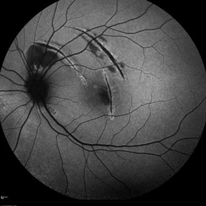
Autofluorescence image of a 39-year-old female patient who sustained blunt ocular trauma resulting in three choroidal ruptures.
Photographer: Ramses Rosales-Diaz, Asociación Para Evitar la Ceguera en México I.A.P., Mexico City
Imaging device: Heidelberg Spectralis
Condition/keywords: blunt trauma, Choroidal Rupture
A project from the American Society of Retina Specialists