-
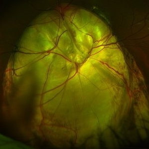
 Vitreomacular Traction
Jun 15 2022 by Zach Seim
Optical Coherence Tomography (OCT) of a 69 year old male with Vitreomacular Traction affecting his right eye. Patient was referred to this office for Choroidal Melanoma in his right eye in May 2021. The patient was treated with Brachytherapy in July 2021 and this OCT was taken at a follow-up appointment in May 2022. Patient's vision was 20/30-2 at the time this OCT was taken. Patient states that his vision was better since his last visit, and that he sees floaters occasionally.
Photographer: Zach Seim
Imaging device: Heidelberg Spectralis
Condition/keywords: heidelberg spectralis, OD, optical coherence tomography (OCT), right eye, subretinal fluid, vitreomacular adhesion, vitreomacular interface disorders, vitreomacular traction (VMT)
-
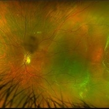
 Choroideremia
Sep 21 2022 by Zach Seim
Ultra-widefield fundus photo of a 74 year old male presenting with severe vision loss beginning at age 55. Patient sought a second opinion with our office and was diagnosed with Choroideremia. Patient denies hearing loss, heart problems, balance issues, polydactyly, kidney problems, and dental problems. Patient reports that nobody in the family had blindness. Choroideremia is an X-linked chorioretinal dystrophy characterized by the diffuse, progressive degeneration of the retinal pigment epithelium (RPE), photoreceptors and choriocapillaris. It is caused by a mutation in the CHM gene.
Photographer: Zach Seim
Imaging device: Optos California
Condition/keywords: choroideremia, hereditary choroidal atrophy, hereditary retinal dystrophy, Optos, pseudocolor, ultra-wide field imaging
-
 Choroideremia
Sep 21 2022 by Zach Seim
Ultra-widefield fundus photo of a 74 year old male presenting with severe vision loss beginning at age 55. Patient sought a second opinion with our office and was diagnosed with Choroideremia. Patient denies hearing loss, heart problems, balance issues, polydactyly, kidney problems, and dental problems. Patient reports that nobody in the family had blindness. Choroideremia is an X-linked chorioretinal dystrophy characterized by the diffuse, progressive degeneration of the retinal pigment epithelium (RPE), photoreceptors and choriocapillaris. It is caused by a mutation in the CHM gene.
Photographer: Zach Seim
Imaging device: Optos California
Condition/keywords: choroideremia, hereditary choroidal atrophy, hereditary retinal dystrophy, left eye, light perception, low vision, Optos, pseudocolor, ultra-wide field imaging
-
 Methotrexate Bubble following Intravitreal Injection for PVR
Sep 21 2022 by Zach Seim
Ultra-widefield fundus photograph of an 81 year old female with a Methotrexate bubble following an Intravitreal Injection for Proliferative Vitreoretinopathy. Patient has been presenting to the office for two week interval Methotrexate injections in her left eye. The image was taken prior to her eighth injection which revealed a residual Methotrexate bubble in her inferior retinal image. This patient was seeing "lots" of floaters, as well as having visual acuity of cc20/400 cc20/200 PH.
Photographer: Zach Seim
Imaging device: OPTOS California
Condition/keywords: bubble, fundus photograph, fundus photography, intravitreal injection, left eye, methotrexate, nasal retina, Optos, proliferative vitreoretinopathy (PVR), pseudocolor, ultra-wide field imaging
-
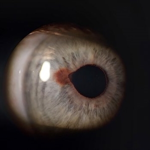
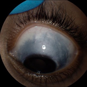
 Iris Coloboma
Feb 22 2023 by Zach Seim
An external image of a 25 year old male with Iris Coloboma, as well as Fundus Coloboma affecting both eyes. Patient's vision at the time of the image was 20/80. Discussed genetic testing as patient reports that he has a child with coloboma and patient agrees. There is a possibility of this finding being syndromic given cornea has small WTW and possibly microphthalmia. Recommended observation without treatment.
Photographer: Zach Seim
Imaging device: Topcon 50DX
Condition/keywords: coloboma, iris, left eye, Topcon
-
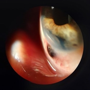
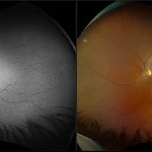
 Fundus Coloboma
Feb 22 2023 by Zach Seim
An ultra-widefield fundus image of a 25 year old male with Fundus Coloboma, as well as Iris Coloboma affecting both eyes. Patient's vision at the time of the image was 20/100-2. Discussed genetic testing as patient reports that he has a child with coloboma and patient agrees. There is a possibility of this finding being syndromic given cornea has small WTW and possibly microphthalmia. The patient has old tractional exudation at edge (abutting fovea). Recommended observation without treatment.
Photographer: Zach Seim
Imaging device: Optos California
Condition/keywords: coloboma, coloboma of optic disc, fundus photograph, Optos, scanning laser ophthalmoscope, ultra-wide field imaging
-
 Macula Off Retinal Detachment
Mar 22 2023 by Zach Seim
An ultra-widefield fundus image of a 65 year old male with a Macula Off Retinal Detachment. Patient's vision at the time of the image was CF at 6 Feet and surgical options were discussed. Fluid-gas exchange was performed without complications.
Photographer: Zach Seim
Imaging device: Optos California
Condition/keywords: left eye, macula off retinal detachment, OPTOS CALIFORNIA, scanning laser ophthalmoscope, ultra-widefield image
-
 Non-Kissing Choroidal Detachment
Apr 3 2023 by Zach Seim
An ultra-widefield fundus image of an 85 year old female with a Choroidal Detachment. Patient's vision at the time of the image was HM and surgery was not recommended.
Photographer: Zach Seim
Imaging device: Optos California
Condition/keywords: choroid, choroidal detachment, left eye, OPTOS CALIFORNIA, ultra-widefield image
-
 Macula Off Retinal Detachment
Jun 23 2023 by Zach Seim
Ultra-widefield fundus photograph of a 42 year old women with a Macula Off Retinal Detachment in her left eye. At the time of this photograph the patient's visual acuity was sc CF at 2 feet.
Photographer: Zach Seim
Imaging device: Optos California
Condition/keywords: left eye, macula off retinal detachment, Optos, OPTOS CALIFORNIA, scanning laser ophthalmoscope, ultra-widefield image
-
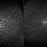
 Neovascularization in Posterior Uveitis
Jul 27 2023 by Zach Seim
An ultra-widefield fluorescein angiogram of a 72 year old male with Posterior Uveitis and Neovascularization affecting the right eye. Patient's vision at the time of the image was Dcc 20/25. Dr. Korot states that the fluorescein angiogram shows patchy leakage throughout both eyes, with peripheral nonperfusion and secondary neovascularization. The patient was asked to get an extensive serological workup in an effort to identify any systemic autoimmune or infectious etiology as the cause for their severe inflammation.
Photographer: Zach Seim
Imaging device: OPTOS California
Condition/keywords: neovascularization (NV), Optos, OPTOS CALIFORNIA, posterior uveitis, right eye, ultra-wide field imaging, ultra-widefield image
-
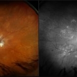
 Coats' Disease
Sep 14 2023 by Zach Seim
An ultra-widefield fluorescein angiogram of a 5 year old male with Coats' Disease affecting the left eye. Patient's vision at the time of the image was Nsc 20/30. Dr. Boss states that the fluorescein angiogram confirms Coats' Disease Stage II.
Photographer: Zach Seim
Imaging device: Optos California
Condition/keywords: Coats' disease, left eye, Optos, OPTOS CALIFORNIA, ultra-wide field imaging
-
 Macular Dystrophy
Oct 25 2023 by Zach Seim
Optos Fundus Autofluorescence of an 84 year old male with Macular Dystrophy. Patient presented with VA of sc CF at 3 feet. Genetic testing was performed to ensure that cause was not genetic.
Photographer: Zach Seim
Imaging device: Optos California
Condition/keywords: Autoflourescence, dystrophy, macular dystrophy, Optos, OPTOS CALIFORNIA, right eye, ultra-wide field imaging
-
 Sturge-Weber Syndrome
Nov 17 2023 by Zach Seim
Topcon photo of a 37 year old female with Sturge-Weber syndrome affecting OU. Patient presents with prominent episcleral vasculature and DCC 20/20 VA OU. Plan to monitor.
Photographer: Zach Seim
Imaging device: Topcon 50DX
Condition/keywords: bilateral, external, external photography, left eye, right eye, Sturge-Weber syndrome, Topcon
-
 Scalloped Choroidal Atrophy
Jan 8 2024 by Zach Seim
An ultra-widefield fluorescein angiogram of a 90 year old female with Scalloped Choroidal Atrophy affecting both eyes. Patient's vision at the time of the image was Dcc 20/40 OD. Genetic test pending.
Photographer: Zach Seim
Imaging device: OPTOS California
Condition/keywords: atrophy, choroidal atrophy, optic nerve, OPTOS CALIFORNIA, retina, right eye, ultra-wide field imaging
-
 Panuveitis
Apr 2 2024 by Zach Seim
Optos Ultra-widefield photo OS of a 59 year old female with Panuveitis OU.
Photographer: Zach Seim
Imaging device: Optos California
Condition/keywords: Optos, OPTOS CALIFORNIA, panuveitis, ULTRA WIDE FIELD, ultra-wide field imaging
-
 Macula Off Retinal Detachment
Jun 25 2024 by Zach Seim
Optos Fundus photo of a 47 year old female with a Macula Off Retinal Detachment right eye, presenting with loss of nasal visual field. Patient's vision at presentation was DCC 20/100-1. Patient was counseled and decided to proceed with surgery.
Photographer: Zach Seim
Imaging device: OPTOS California
Condition/keywords: macula off retinal detachment, Optos, OPTOS CALIFORNIA, right eye
-
 Iris Nevus
Jul 3 2024 by Zach Seim
Slit Lamp Photograph of an 88 year old man with an Iris Nevus. Patient presented with DCC 20/60+1. Plan to monitor.
Photographer: Zach Seim
Imaging device: Slit Lamp photography with Samsung Galaxy 7
Condition/keywords: iris, iris nevus, nevus, right eye, slit lamp photo, slit lamp photography
-
 Proliferative Diabetic Retinopathy
Jul 5 2024 by Zach Seim
Fluorescein Angiogram of a 44 year-old female with PDR.
Photographer: Zach Seim
Imaging device: Optos California
Condition/keywords: FA early phase, Optos, OPTOS CALIFORNIA, proliferative diabetic retinopathy (PDR)
-
 Asteroid Hyalosis
Jul 5 2024 by Zach Seim
Optos fundus photo of an 88 year old male with Asteroid Hyalosis OS. VA Dsc 20/30-2 upon presentation.
Photographer: Zach Seim
Imaging device: Optos California
Condition/keywords: ASTEROID, asteroid hyalosis, optos, OPTOS CALIFORNIA
-
 Adult Onset Coats' Disease
Jul 5 2024 by Zach Seim
FA/ICG of a 64 year old female with Adult Onset Coats' Disease. VA DCC CF@3 feet upon presentation. Therapy options discussed extensively.
Photographer: Zach Seim
Imaging device: Optos California
Condition/keywords: Coats' disease, FA, indocyanine green (ICG) angiography, Optos, OPTOS CALIFORNIA
-
 Congenital Nuclear Cataract
Jul 5 2024 by Zach Seim
This is a slit-lamp photograph of a 10 year old female with a congenital nuclear cataract OD. Patient presented with VA Dsc 20/200. Patient was counseled on surgical options.
Photographer: Zach Seim
Imaging device: Slit Lamp Photography on Samsung Galaxy 7
Condition/keywords: cataract, congenital cataract, nuclear sclerosis, right eye, slit lamp photo
-
 Posterior Uveitis
Jul 5 2024 by Zach Seim
FA/ICG OS of a 39 year old female with Posterior Uveitis. VA at time of photos was Dsc 20/20-1.
Photographer: Zach Seim
Imaging device: Optos California
Condition/keywords: FA, indocyanine green (ICG) angiography, Optos, OPTOS CALIFORNIA, posterior uveitis
-
 Scleral Ectasia Post Radiation for Iris Melanoma
Jul 5 2024 by Zach Seim
Slit-Lamp Photograph of a 52 year old male with Scleral Ectasia post radiation for Iris Melanoma.
Photographer: Zach Seim
Imaging device: Slit Lamp Photography on Samsung Galaxy 7
Condition/keywords: Iris, iris melanoma, scleral ectasia, slit lamp photo, slit lamp photography
-
 Central Retinal Vein Occlusion with Macular Edema OS
Jul 5 2024 by Zach Seim
Optos fundus photograph of a Central Retinal Vein Occlusion in a 20 year old male. Vision at presentation was Dsc 20/25-1.
Photographer: Zach Seim
Imaging device: Optos California
Condition/keywords: central retinal vein occlusion (CRVO), left eye, macular edema, Optos, OPTOS CALIFORNIA
-
 Disseminated Retinitis and Retinochoroiditis, Pigment Epitheliopathy OS
Jul 5 2024 by Zach Seim
Optos Fluorescein Angiogram of a 94 year old female with Disseminated Retinitis and Retinochoroiditis, Pigment Epitheliopathy OS.
Photographer: Zach Seim
Imaging device: Optos California
Condition/keywords: left eye, OPTOS CALIFORNIA, retinitis, retinochoroiditis
-
 Choroidal Detachment OS
Jul 5 2024 by Zach Seim
Optos Fundus Photograph of a Choroidal Detachment OS in a 75 year old male. VA at presentation was DCC HM.
Photographer: Zach Seim
Imaging device: Optos California
Condition/keywords: choroidal detachment, choroidal mass, left eye, optos, OPTOS CALIFORNIA
-
 Oculodermal Melanocytosis OS
Jul 5 2024 by Zach Seim
Topcon photograph of a 13 year old with Oculodermal Melanocytosis OS.
Photographer: Zach Seim
Imaging device: Topcon 50DX
Condition/keywords: left, left eye, Oculodermal Melanocytosis, OS, Topcon
-
 Endophthalmitis
Jul 5 2024 by Zach Seim
OCT of a 59 year old male with Fungal Endophthalmitis. The fungus can be seen breaking through in the OCT.
Photographer: Zach Seim
Imaging device: Heidelberg OCT
Condition/keywords: endophthalmitis, fungal, OCT
-
 Endophthalmitis
Jul 5 2024 by Zach Seim
Optos Fundus Photograph of a 59 year old male with Fungal Endophthalmitis. Patient presented with VA Dsc 20/70.
Photographer: Zach Seim
Imaging device: Optos California
Condition/keywords: endophthalmitis, fundus photograph, fungal, optos, OPTOS CALIFORNIA
-
 Macula On Retinal Detachment
Jul 5 2024 by Zach Seim
This is an Optos fundus photo of a 67 year old female with a Macula On Retinal Detachment. Patient presented with VA DCC 20/40-1.
Photographer: Zach Seim
Imaging device: Optos California
Condition/keywords: macula on, Optos, OPTOS CALIFORNIA, right eye
-
 Posterior Uveitis
Jul 5 2024 by Zach Seim
Fluorescein Angiogram of a 73 year old Male with Posterior Uveitis OS. Patient presented with VA of DCC 20/80-1 OS at this visit.
Photographer: Zach Seim
Imaging device: Optos California
Condition/keywords: fa, Optos, OPTOS CALIFORNIA, posterior uveitis
-
 Sclerochoroidal Calcification
Jul 5 2024 by Zach Seim
Optos fundus photo of an 81 year old female with Sclerochoroidal Calcification. Patient's VA at presentation was DCC 20/70-1.
Photographer: Zach Seim
Imaging device: Optos California
Condition/keywords: optos, OPTOS CALIFORNIA, sclerochoroidal calcification
A project from the American Society of Retina Specialists