-
 Gyrate Atrophy of the Choroid and Retina
May 1 2019 by Anmol Naik
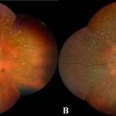
A 34-year-old Indian male presented with gradual progressive bilateral diminution of peripheral vision since 6 years. His best corrected visual acuity was 6/60, N36 in right eye and 6/9, N6 in left. Wide-field fundus imaging demonstrated scalloped areas of chorioretinal atrophy with well-defined margins. His plasma ornithine levels were elevated.at 203.9 nmol/ml. Based on the typical features, a diagnosis of gyrate atrophy was made.
Photographer: Anmol Naik, Sankara Nethralaya, Chennai, India
Imaging device: Zeiss CLARUS 500
Condition/keywords: chorioretinal atrophy, gyrate atrophy
-
 Pseudoexfoliation Deposit on Intraocular Lens
Jun 12 2021 by Anmol Naik
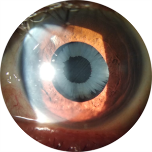
A 64-year-old Indian farmer presented with complaint of diminution of vision. Observed here is the deposition of pseudoexfoliation material on the intraocular lens implant in a typical florette pattern. Glaucoma is one of the known and vision threatening complications in such patients.
Photographer: Dr. Anmol Naik, MS, Nakshatra Superspeciality Eye Hospital, Pune, India.
Imaging device: Appasamy Slit Lamp Imaging system - 11 3S L, Appasamy Associates, Chennai, India.
Condition/keywords: pseudoexfoliation syndrome
A project from the American Society of Retina Specialists