-
 Slide 12-1
Feb 26 2019 by Lancaster Course in Ophthalmology
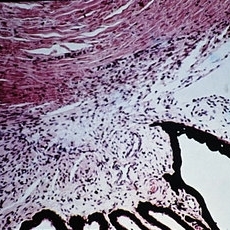
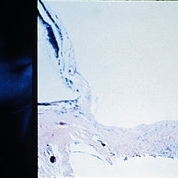
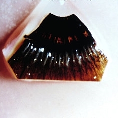
Congenital glaucoma. Anterior chamber angle from a 2-year-old child with congenital glaucoma is cut tangentially and resembles the configuration seen normally in a fetus (H&E xlOl).
Condition/keywords: congenital glaucoma
-
 Slide 12-2
Feb 27 2019 by Lancaster Course in Ophthalmology
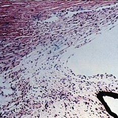
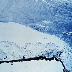
Congenital glaucoma. Meridional microscopic section from the same child shows a more normal appearance (H&E xlOl).
Condition/keywords: congenital glaucoma
-
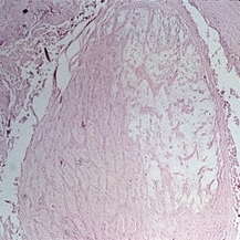
 Slide 12-3
Feb 27 2019 by Lancaster Course in Ophthalmology
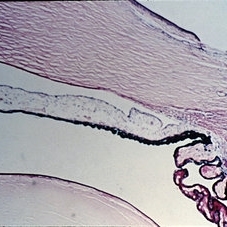
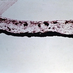
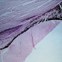
Congenital glaucoma. Anterior half of eye stretched and enlarged. Stretching of the limbal area and sclera over the anterior portion of the ciliary body (intercalary area) is called intercalary ectasia (H&E x3).
Condition/keywords: congenital glaucoma
-
 Slide 12-4
Feb 27 2019 by Lancaster Course in Ophthalmology
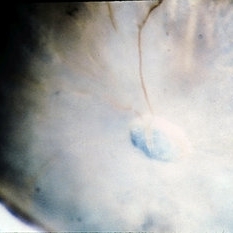
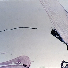
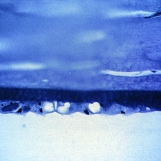
Congenital glaucoma. The ruptured end of a Descemet's membrane has healed over so that the scroll-like extension of the membrane hanging into the anterior chamber is covered completely by endothelium (H&E xlOl).
Condition/keywords: congenital glaucoma , descemet's membrane
-
 Slide 12-5
Feb 27 2019 by Lancaster Course in Ophthalmology
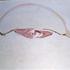
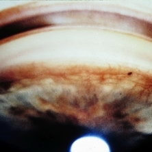
Congenital glaucoma. Deep glaucoma cup of the optic disk is shown macroscopically in an eye from a 7-year-old with congenital glaucoma.
Condition/keywords: congenital glaucoma
-
 Slide 12-6
Feb 27 2019 by Lancaster Course in Ophthalmology
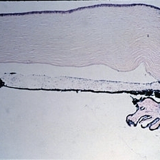
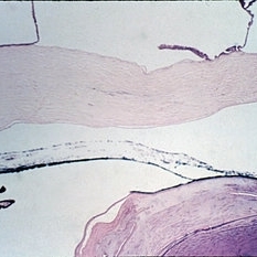
Primary closed-angle glaucoma. The anterior chamber is shallow and its angle is narrowed with the iris root in contact with the posterior portion of the trabecular meshwork (PASx28).
Condition/keywords: primary angle-closure glaucoma
-
 Slide 12-7
Feb 27 2019 by Lancaster Course in Ophthalmology
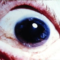
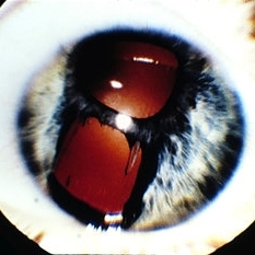
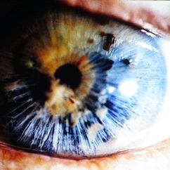
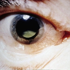
Primary closed-angle glaucoma. The clinical triad of irregular pupil, segmental iris atrophy, and glaukomflecken is seen.
Condition/keywords: glaukomflecken, primary angle-closure glaucoma
-
 Slide 12-8
Feb 27 2019 by Lancaster Course in Ophthalmology
A necrotic iris is shown, with loss of stroma as well as dilator and sphincter muscles (H&E x21).
Condition/keywords: retinal necrosis
-
 Slide 12-9
Feb 27 2019 by Lancaster Course in Ophthalmology
Primary open-angle glaucoma. The macroscopic (left) and histologic (right) appearance of a deeply cupped optic nervehead is shown (right view, PAS x16).
Condition/keywords: primary open-angle glaucoma
-
 Slide 12-10
Feb 27 2019 by Lancaster Course in Ophthalmology
Essential iris atrophy. Pupil (above) and full-thickness iris hole show red reflex.
Condition/keywords: iris atrophy
-
 Slide 12-11
Feb 27 2019 by Lancaster Course in Ophthalmology
Essential iris atrophy. Anterior chamber angle closed by a peripheral anterior synechia (H&E x16).
Condition/keywords: iris atrophy
-
 Slide 12-12
Feb 27 2019 by Lancaster Course in Ophthalmology
Essential iris atrophy. An area showing a full-thickness iris hole (H&Ex21).
Condition/keywords: iris atrophy
-
 Slide 12-13
Feb 27 2019 by Lancaster Course in Ophthalmology
Essential iris atrophy. An area showing a full-thickness iris hole (H&Ex21).
Condition/keywords: iris atrophy
-
 Slide 12-14
Feb 27 2019 by Lancaster Course in Ophthalmology
lridoschisis. Clinical appearance of two different cases. Iris stroma is loosened and resembles spaghetti.
Condition/keywords: iridoschisis
-
 Slide 12-15
Feb 27 2019 by Lancaster Course in Ophthalmology
lridoschisis. Macroscopic appearance of the same cases.
Condition/keywords: iridoschisis
-
 Slide 12-16
Feb 27 2019 by Lancaster Course in Ophthalmology
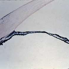
lridoschisis. The large space present abnormally between the anterior and posterior iris stromal fibers. Note the lifting off of the corneal epithelium to form a bullous keratopathy (H&E x16).
Condition/keywords: epithelium, iridoschisis, keratopathy
-
 Slide 12-17
Feb 27 2019 by Lancaster Course in Ophthalmology
Rubeosis iridis. Vessels climbing over the anterior chamber angle have produced peripheral anterior synechiae.
Condition/keywords: rubeosis
-
 Slide 12-18
Feb 27 2019 by Lancaster Course in Ophthalmology
Rubeosis iridis. Fibrovascular tissue is present abnormally anterior to the anterior border layer of the iris. Shrinkage of such abnormal tissue results frequently in ectropion uveae (H&E xlOl).
Condition/keywords: fibrovascular tissue, rubeosis
-
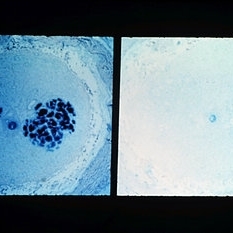
 Slide 12-19
Feb 27 2019 by Lancaster Course in Ophthalmology
Pigmentary glaucoma. A Krukenberg spindle results from the phagocytosis of pigment by the corneal endothelium (1.5pm section, PD x200).
Condition/keywords: glaucoma, Krukenberg's spindle
-
 Slide 12-20
Feb 27 2019 by Lancaster Course in Ophthalmology
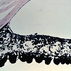
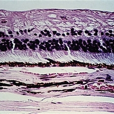
Pigmentary glaucoma. Pigment is liberated from the posterior iris pigment epithelium and is then carried to the anterior chamber where it is phagocytosed by the endothelium of the cornea and the trabecular meshwork (H&E x40).
Condition/keywords: endothelium, glaucoma, phagocytosed
-
 Slide 12-21
Feb 27 2019 by Lancaster Course in Ophthalmology
Pigmentary glaucoma. Macroscopic appearance of the back of the iris. Note that the main loss of iris pigment epithelium occurs where the middle and peripheral thirds of the iris meet.
Condition/keywords: glaucoma
-
 Slide 12-22
Feb 27 2019 by Lancaster Course in Ophthalmology
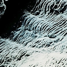
Pigmentary glaucoma. Scanning electron microscopic appearance of the back of the iris, showing loss (flattening) of the circumferential ridges of the posterior iris at the sites of involvement (SEM x50).
Condition/keywords: glaucoma
-
 Slide 12-23
Feb 27 2019 by Lancaster Course in Ophthalmology
Pigmentary glaucoma. Necrosis of the iris pigment epithelium, macrophages filled with pigment in the iris stroma, and atrophy and hyperplasia of the iris dilator muscle are present (H&E x101).
Condition/keywords: glaucoma, hyperplasia, retinal necrosis
-
 Slide 12-24
Feb 27 2019 by Lancaster Course in Ophthalmology
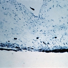
Sequelae. Basal layer of the corneal epithelium is pale and swollen by edema fluid. Further accumulation of edema fluid will lift the epithelium off of Bowman's membrane, resulting in bullous keratopathy (see Slide 12-16) (Masson's tri chromex250).
Condition/keywords: edema, epithelium, sequelae
-
 Slide 12-25
Feb 27 2019 by Lancaster Course in Ophthalmology
Sequelae. The anterior chamber is deep, and the pupillary iris shows ectropion uveae resulting from shrinkage of new fibrovascular tissue (rubeosis iridis) on the anterior surface of the iris. The lens is dislocated posteriorly into the vitreous. All changes are the result of blunt trauma.
Condition/keywords: ectropion uveae, rubeosis, sequelae
-
 Slide 12-26
Feb 27 2019 by Lancaster Course in Ophthalmology
Sequelae. Massive peripheral anterior synechia and ectropion uveae have resulted from blunt trauma. Note the postcontusion deformity of the anterior chamber angle (H&E x40).
Condition/keywords: anterior synechiae, ectropion uveae, sequelae
-
 Slide 12-27
Feb 27 2019 by Lancaster Course in Ophthalmology
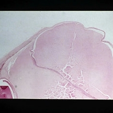
Sequelae. Increased intraocular pressure has resulted in stretching and thinning of the sclera in the equatorial region, resulting in an equatorial staphyloma (H&E x3).
Condition/keywords: sclera, sequelae, staphyloma
-
 Slide 12-28
Feb 27 2019 by Lancaster Course in Ophthalmology
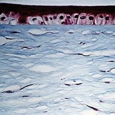
Sequelae. The inner retinal layers are atrophic in this case of long standing glaucoma (H&E x101).
Condition/keywords: sequelae
-
 Slide 12-29
Feb 27 2019 by Lancaster Course in Ophthalmology
Sequelae. Cavernous degeneration of the optic nerve (Schnabel's cavernous atrophy) shows cystic spaces which appear clear in hematoxylin and eosin stained sections (H&E x21).
Condition/keywords: optic atrophy, sequelae
-
 Slide 12-30
Feb 27 2019 by Lancaster Course in Ophthalmology
Sequelae. Stains for acid mucopolysaccharides (left) show spaces to contain AMP. The AMP disappears if the tissue on the slide is first digested with hyaluronidase (right), showing that the material is hyaluronidase-sensitive AMP, presumably hyaluronic acid (left view, AMPxl6; right view, hyaluronidase-AMPx16).
Condition/keywords: sequelae
A project from the American Society of Retina Specialists