-
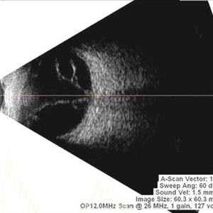 Multiple Retinal Cysts Associated With Chronic Retinal Detachment
Sep 24 2018 by samarth mishra
Patient presented with a diminution of vision in left eye since few months. On B-scan ultrasonography multiple retinal cysts with a total retinal detachment were noted.
Photographer: Aditya Birla Sankara Nethralaya, West Bengal , Kolkata , India
Condition/keywords: B scan ultrasound, chronic retinal detachment, intraretinal cyst, retinal cyst
-
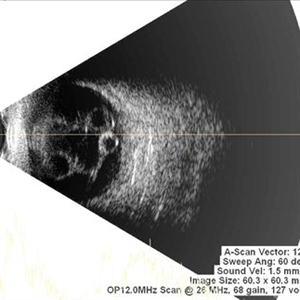 Multiple Retinal Cysts Associated With Chronic Retinal Detachment
Sep 24 2018 by samarth mishra
Patient presented with a diminution of vision in left eye since few months. On B-scan ultrasonography multiple retinal cysts with a total retinal detachment were noted.
Photographer: Aditya Birla Sankara Nethralaya, West Bengal , Kolkata , India
Condition/keywords: B scan ultrasound, chronic retinal detachment, intraretinal cyst, retinal cyst
-
 Retinal Detachment Sparing Fovea By Microns
Sep 24 2018 by samarth mishra
A 29-year-old young female presented with complaint of blurring of vision in the right eye since one year. Best corrected visual acuity was 20/40. On routine examination inferior retinal detachment was noted. Optical coherence tomography (OCT) showed the retinal detachment sparing the fovea by few microns.
Photographer: Aditya Birla Sankara Nethralaya, Kolkata , West Bengal , India
Condition/keywords: color fundus photograph, multicolor, optical coherence tomography (OCT)
-
 Retinal Detachment Sparing Fovea By Microns
Sep 24 2018 by samarth mishra
A 29-year-old young female presented with complain of blurring of vision in the right eye since one year. Best corrected visual acuity was 20/40. On routine examination inferior retinal detachment was noted. Optical coherence tomography (OCT) showed the retinal detachment sparing the fovea by few microns.
Photographer: Aditya Birla Sankara Nethralaya, Kolkata , West Bengal , India
Condition/keywords: color fundus photograph, multicolor, optical coherence tomography (OCT)
-
 Multicolor Imaging of Bilateral Branch Retinal Vein Occlusion
Sep 25 2018 by samarth mishra
A 40-year-old female presented with complains of blurring of vision since past 1 week. Patient had a history of hypertension. On routine examination bilateral branch retinal vein occlusion was noted. Visual acuity at presentation was 6/9 and 6/15 in the right and left eye respectively. Multicolor composite imaging shows the hemorrhage as red and the retinal thickening as greenish hue. She was managed with anti vascular endothelial growth factor in both eyes.
Photographer: Aditya Birla Sankara Nethralaya, Kolkata, West Bengal , India
Condition/keywords: branch retinal vein occlusion (BRVO), Heidelburg Spectralis, multicolor, optical coherence tomography (OCT)
-
 Retinal Detachment Sparing Fovea By Microns
Sep 25 2018 by samarth mishra
A 29-year-old young female presented with complain of blurring of vision in the right eye since one year. Best corrected visual acuity was 20/40. On routine examination inferior retinal detachment was noted. Optical coherence tomography (OCT) showed the retinal detachment sparing the fovea by few microns.
Photographer: Aditya Birla Sankara Nethralaya, Kolkata, West Bengal , India
Condition/keywords: color fundus photograph, infrared image, multicolor, optical coherence tomography (OCT), red-free
-
 Multicolor Imaging in Diabetic Retinopathy
Sep 25 2018 by samarth mishra
A 60-year-old male presented with a history of blurring of vision since many months. He had a history of diabetes since last 8 years. On routine examination proliferative diabetic retinopathy with diabetic macular edema was noted. Fundus fluorescein angiography showed neovascularization elsewhere. Hard exudates can be seen as greenish yellow dots all over the posterior pole in multicolor imaging. Retinal hemorrhage can be seen as dark red.
Photographer: Aditya Birla Sankara Nethralaya, Kolkata, West Bengal , India
Condition/keywords: diabetic retinopathy, Heidelburg Spectralis, multicolor, optical coherence tomography (OCT)
-
 Stage 3 Macular Hole With Operculum
Sep 25 2018 by samarth mishra
Stage 3 macular hole with operculum.
Photographer: Aditya Birla Sankara Nethralaya, Kolkata, West Bengal , India and Sankara Nethralaya, chennai , India
Condition/keywords: full thickness macular hole, macular hole, optical coherence tomography (OCT)
A project from the American Society of Retina Specialists