-
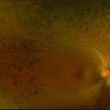 Retinitis Pigmentosa - Color OD
Jun 18 2018 by Hosam Attia, MD
Pseudo-color ultra-wide fundus photograph of a 38-year-old African American female with degenerative myopia and Unilateral RP variant, depicting extensive mid-peripheral bone spicules, extending further into the periphery, with relative sparing of the macula OD. VF 30-V showed severe peripheral constriction OD, enlarged BS OS and OCT showed severe ellipsoid zone degeneration with saucerization and cystoid macular degeneration with no obvious late macular leakage on FA (Both, not shown)
Imaging device: Optos California
Condition/keywords: bone spicule, peripheral bone spicules, retinitis pigmentosa
-
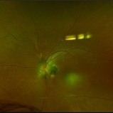 Retinitis Pigmentosa - Color OS
Jun 18 2018 by Hosam Attia, MD
38-year-old female with unilateral retinitis pigmentosa.
Imaging device: Optos California
Condition/keywords: bone spicule, peripheral bone spicules, retinitis pigmentosa
-
 Retinitis Pigmentosa - Autofluorescence OD
Jun 18 2018 by Hosam Attia, MD
Ultra-wide fundus auto-fluorescence photograph of a 38-year-old African, American female with degenerative myopia, unilateral RP variant, depicting extensive mid-peripheral bone spicules hypo-autofluorescence, extending further into the periphery w/ relative sparing of the macula OD VF 30-V showed severe peripheral constriction OD, enlarged BS OS and OCT showed severe ellipsoid zone degeneration with saucerization and cystoid macular degeneration with no obvious late macular leakage on FA (Both, not shown)
Imaging device: Optos California
Condition/keywords: bone spicule, peripheral bone spicules, retinitis pigmentosa
-
 Retinitis Pigmentosa - Autofluorescence OS
Jun 18 2018 by Hosam Attia, MD
Retinitis Pigmentosa
Imaging device: Optos California
Condition/keywords: bone spicule, peripheral bone spicules, retinitis pigmentosa
-
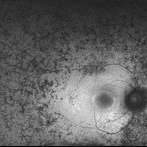
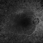 Retinitis Pigmentosa - Fluorescein Angiogram OD
Jun 18 2018 by Hosam Attia, MD
Ultra-wide fluorescein angiogram of a 38-year-old African, American female with degenerative myopia, Unilateral RP variant, depicting abnormal fluorescence pattern with extensive mid-peripheral bone spicules hypofluorescence, extending further into the periphery w/ relative sparing of the macula OD. VF 30-V showed severe peripheral constriction OD, enlarged BS OS & OCT showed severe ellipsoid zone degeneration with saucerization and cystoid macular degeneration w/ No obvious late macular leakage on FA (Both, not shown)
Imaging device: Optos California
Condition/keywords: bone spicule, peripheral bone spicules, retinitis pigmentosa
-
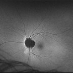
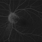 Retinitis Pigmentosa - Fluorescein Angiogram OS
Jun 18 2018 by Hosam Attia, MD
38-year-old African American female with unilateral retinitis pigmentosa.
Imaging device: Optos California
Condition/keywords: bone spicule, peripheral bone spicules, retinitis pigmentosa
A project from the American Society of Retina Specialists