-
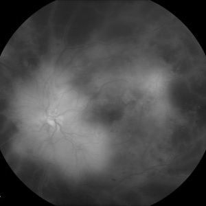
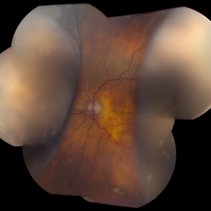
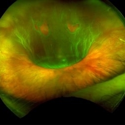
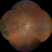
 Optic Nerve Head Drusen With Idiopathic CNV
Feb 17 2017 by Kristen Wagner
22-year-old female fundus photograph of a right eye with Optic Nerve Drusen with Idiopathic CNV.
Photographer: Kristen Wagner, COT, OSC Ophthalmic Photographer, Tennessee Retina, Nashville TN
Condition/keywords: choroidal neovascularization (CNV), drusen of optic disc, optic disc drusen
-
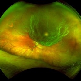
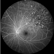
 Severe NVD Leakage During a Fluorescein Angiogram
Mar 26 2018 by Kristen Wagner
Fluorescein angiogram image of the disc of a left eye showing leakage (NVD). Patient is a young woman with uncontrolled Diabetes Type II with severe neovascularization of the disc (NVD) and PDR.
Photographer: Kristen Wagner, COT, OSC
Condition/keywords: diabetes, fluorescein leakage, neovascularization (NV), neovascularization of the disc (NVD), optic disc, optic nerve, proliferative diabetic retinopathy (PDR)
-
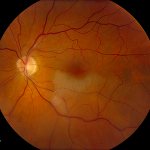
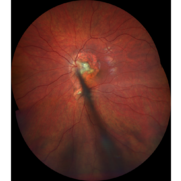
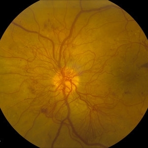 Severe NVD
Mar 26 2018 by Kristen Wagner
Fundus photograph of a young woman with uncontrolled Diabetes Type II with severe neovascularization of the disc (NVD) and PDR.
Photographer: Kristen Wagner, COT, OSC
Condition/keywords: diabetes, neovascularization (NV), neovascularization of the disc (NVD), optic disc, optic nerve, proliferative diabetic retinopathy (PDR)
-
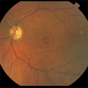
 Retinoschesis Inferotemporal
Dec 2 2019 by Kristen Wagner
Retinoschisis inferotemporal of a young patient. Vision was 20/20.
Photographer: Kristen Wagner, COT, OSC, Ophthalmic Photographer, Tennessee Retina
Condition/keywords: retinoschisis
-
 Subretinal Hemorrhage
Dec 2 2019 by Kristen Wagner
Fundus photo of a very large subretinal hemorrhage.
Photographer: Kristen Wagner, COT, OSC, Ophthalmic Photographer, Tennessee Retina
Condition/keywords: hemorrhage
-
 Choroidal Detachment OS
Dec 2 2019 by Kristen Wagner
Choroidal Detachment of the left eye. Patient's vision was Hand Motion best corrected.
Photographer: Kristen Wagner, COT, OSC, Ophthalmic Photographer, Tennessee Retina
Condition/keywords: choroidal detachment
-
 Branch Retinal Artery Occlusion
Dec 2 2019 by Kristen Wagner
Fundus Photo of a left eye with a branch retinal artery occlusion. Vision was DVA cc 20/40.
Photographer: Kristen Wagner, COT, OSC, Ophthalmic Photographer, Tennessee Retina
Condition/keywords: branch retinal artery occlusion (BRAO)
-
 Retinal Detachment with Multiple Retinal Tears
Jan 13 2021 by Kristen Wagner
Optos image of a male with a retinal detachment and multiple retinal tears.
Photographer: Kristen Wagner, COT Tennessee Retina Nashville TN
Condition/keywords: detachment, retinal tear with detachment
-
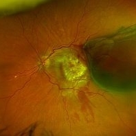
 Large Macular Hole
Jan 13 2021 by Kristen Wagner
Topcon image of a female with a large chronic macular hole in the left eye.
Photographer: Kristen Wagner, COT Tennessee Retina Nashville TN
Condition/keywords: macular hole
-
 Retinal Detachment with Multiple Retinal Tears
Jan 13 2021 by Kristen Wagner
Optos image of a male with a macular off retinal detachment and multiple retinal tears. This patient had detachment in both eyes.
Photographer: Kristen Wagner, COT Tennessee Retina Nashville TN
Condition/keywords: retinal detachment of the macula, retinal tear, retinal tear with detachment
-
 Branch Retinal Vein Occlusion
Jun 18 2021 by Kristen Wagner
Angiogram of a super temporal branch retinal vein occlusion on a 51-year-old male.
Photographer: Kristen Wagner, COT Tennessee Retina Nashville TN
Imaging device: Clarus
Condition/keywords: branch retinal vein occlusion (BRVO), macular branch retinal vein occlusion (BRVO)
-
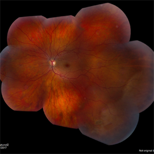
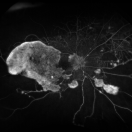
 Nerve Stalk
Jun 18 2021 by Kristen Wagner
Fundus photograph of a hyperplastic primary vitreous, nerve stalk, and choroidal lesion of a 7-year-old male.
Photographer: Kristen Wagner, COT Tennessee Retina Nashville TN
Imaging device: Clarus
Condition/keywords: choroidal lesions, nerve, vitreous
-
 Severe Diabetic Retinopathy and NVE with Macular Hole
Jun 18 2021 by Kristen Wagner
Fundus photo of severe PDR with NVE and nonperfusion. The patient also has a macular hole.
Photographer: Kristen Wagner, COT Tennessee Retina Nashville TN
Imaging device: Optos
Condition/keywords: diabetic macular edema, macular hole, neovascularization elsewhere (NVE), proliferative diabetic retinopathy (PDR)
-
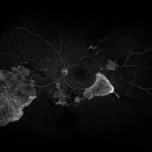
 Severe Diabetic Retinopathy, Nonperfusion and NVE
Jun 18 2021 by Kristen Wagner
Optos angiogram of severe diabetic retinopathy, nonperfusion and NVE.
Photographer: Kristen Wagner, COT Tennessee Retina Nashville TN
Imaging device: Optos
Condition/keywords: diabetic macular edema, neovascularization elsewhere (NVE), nonperfusion diabetic retinopathy, proliferative diabetic retinopathy (PDR)
-
 Sickle Cell
Jul 6 2021 by Kristen Wagner
Fundus photo on the Clarus who is diagnosed with sickle cell retinopathy and has had laser treatment.
Photographer: Kristen Wagner COT, Tennessee Retina, Nashville TN
Condition/keywords: sickle cell retinopathy
-
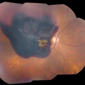
 Hemorrhagic Choroidal Detachment
Jul 6 2021 by Kristen Wagner
Hemorrhagic choroidal detachment with overlying retinal detachment, neovascular AMD with inactive scar.
Photographer: Kristen Wagner COT, Tennessee Retina, Nashville TN
Imaging device: optos
Condition/keywords: choroidal detachment, hemorrhage, wet age-related macular degeneration (wet AMD)
-
 B-scan Ultrasound of Choroidal Melanoma with Serous Retinal Detachment
Sep 5 2025 by Kristen Wagner
B-scan ultrasound of a choriodal melanoma with serous retinal detachment.
Photographer: Kristen Wagner, COT Tennessee Retina
Condition/keywords: B scan ultrasound, Choroidal melanoma, serous retinal detachment
-
 X-Linked Juvenile Retinoschisis
Nov 5 2025 by Kristen Wagner
Fundus photograph of a 24 year old male patient who has X-Linked Retinoschisis (XLRS). Findings include Mild to moderate diffuse maculoschisis OD with vitreous veils. Discussed mutations with RS1 gene.
Photographer: Kristen Cross, COT Tennessee Retina
Imaging device: Optos
Condition/keywords: juvenile retinoschisis, maculoschisis, retinoschisis, virreous veils
A project from the American Society of Retina Specialists