Note the relatively flat, peripapillary, yellow white tumor with overlying retinal pigment epithelial hypertrophy and atrophy. Also note the scalloped peripheral edges.
-
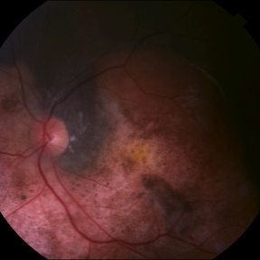 Choroidal Osteoma
Aug 29 2014 by Paul T. Finger, MD, FACS
Note the relatively flat, yellow-white tumor with overlying clusters of RPE hypertrophy and scalloped edges. This choroidal osteoma also had CNV that responded to photodynamic therapy.
Imaging device: Topcon
Condition/keywords: choroidal osteoma
-
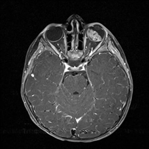 MRI: The Preferred Method to Image the Brain and Orbit of Children with Retinoblastoma
Oct 2 2015 by Paul T. Finger, MD, FACS
MRI used to evaluate a child with retinoblastoma. Note the white tumor on the T1 weighted image. There is no evidence of extrascleral or intramural invasion nor PNET.
Imaging device: MRI
Condition/keywords: MRI, retinoblastoma
-
 Retinoblastoma with Vitreous Seeding
Oct 2 2015 by Paul T. Finger, MD, FACS
Exophytic retinoblastoma shedding clumps of tumor (seeds).
Photographer: Anonymous
Condition/keywords: retinoblastoma, vitreous, vitreous seeding
-
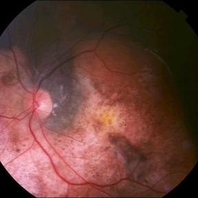 Peripapillary Choroidal Osteoma
Oct 2 2015 by Paul T. Finger, MD, FACS
Note the lightly albeit variably pigmented peripapillary tumor. It is relatively flat, with overlying evidence of RPE hypertrophy. Note the scalloped edges. CNV is not seen in this case.
Photographer: anonymous
Condition/keywords: choroidal osteoma