 Initializing download.
-
 By Paul T. Finger, MD, FACS
The New York Eye Cancer Center
Co-author(s): Paul T Finger, MD, The New York Eye Cancer Center, New York, NY, USA - Uploaded on Oct 2, 2015.
- Last modified by Caroline Bozell on Oct 2, 2015.
- Rating
- Appears in
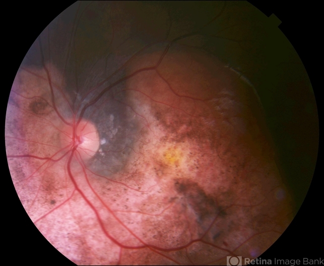
- Choroidal Osteoma
- Condition/keywords
- choroidal osteoma
- Photographer
- anonymous
- Imaging device
- Fundus camera
- Description
- Note the lightly albeit variably pigmented peripapillary tumor. It is relatively flat, with overlying evidence of RPE hypertrophy. Note the scalloped edges. CNV is not seen in this case.


---thumb.jpg/image-square;max$79,0.ImageHandler "Choroidal osteoma case 1 no 2")
---thumb.jpg/image-square;max$79,0.ImageHandler "Choroidal osteoma case 1 no 3")