-
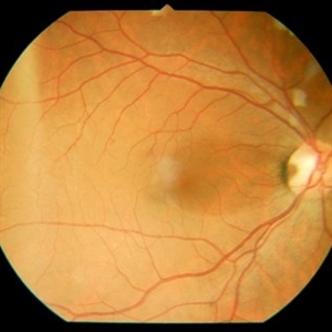
 Color Fundus Photograph of Macular Infarction Secondary to Subonjunctival Gentamicin Injection
May 16 2014 by Arwa Azmeh, MD, PhD
A 20-year-old male suffered from diplopia since age one. He was diagnosed to have acquired fourth nerve palsy in his left eye. VA at time of diagnosis was 20/20 in OU and Fundus exam was WNL in OU. His history revealed no other complaints. 3 days ago he underwent left superior oblique tucking for relief of his diplopia.The surgery was uneventful and at the end of surgery subconjunctival gentamicin was injected. Immediately following surgery his VA in OS decreased from 20/20 to complete loss of central vision and sensation of HM from the periphery. He was referred to us 3 days after surgery. At time of referral fundus exam of his left eye revealed macular infarction with cherry red spot appearance with few retinal hemorrhages, mild optic disc edema and CWS surrounding optic disc. Peripheral retina had normal color and appearance. The vitreous was clear. Anterior segment was quiet. IOP was WNL. Macular OCT was consistent with macular infarction. FA revealed delay in central retinal artery filling as fluorescein started to appear in the arteries at the level of the optic disc at 28 sec, and in the retinal veins at 38 sec. Macular area remained to be non-perfused throughout the whole FA. In late phases staining of blood vessels walls was noticed. The "wipe out" of large vessels and capillaries persisted in the central area. OCT through foveal area showed diffuse thickening of the retina with severe elevation in the fovea, reduced backscattering from the outer layers of the retina and enhanced reflectivity from the inner retina, due to ischemia. Complete blood count and cardiovascular study were WNL. The final diagnosis was macular infarction secondary to subconjunctival gentamicin injection.
Imaging device: OCT
Condition/keywords: macular infarction, subconjunctival gentamicin
-
 Early-FA-phase-of-macular-infarction-secondary-to-subconjunctival-gentamycin-injection
May 16 2014 by Arwa Azmeh, MD, PhD
A 20-year-old male suffered from diplopia since age one. He was diagnosed to have acquired fourth nerve palsy in his left eye. VA at time of diagnosis was 20/20 in OU and Fundus exam was WNL in OU. His history revealed no other complaints. 3 days ago he underwent left superior oblique tucking for relief of his diplopia.The surgery was uneventful and at the end of surgery subconjunctival gentamicin was injected. Immediately following surgery his VA in OS decreased from 20/20 to complete loss of central vision and sensation of HM from the periphery. He was referred to us 3 days after surgery. At time of referral fundus exam of his left eye revealed macular infarction with cherry red spot appearance with few retinal hemorrhages, mild optic disc edema and CWS surrounding optic disc. Peripheral retina had normal color and appearance. The vitreous was clear. Anterior segment was quiet. IOP was WNL. Macular OCT was consistent with macular infarction. FA revealed delay in central retinal artery filling as fluorescein started to appear in the arteries at the level of the optic disc at 28 sec, and in the retinal veins at 38 sec. Macular area remained to be non-perfused throughout the whole FA. In late phases staining of blood vessels walls was noticed. The "wipe out" of large vessels and capillaries persisted in the central area. OCT through foveal area showed diffuse thickening of the retina with severe elevation in the fovea, reduced backscattering from the outer layers of the retina and enhanced reflectivity from the inner retina, due to ischemia. Complete blood count and cardiovascular study were WNL. The final diagnosis was macular infarction secondary to subconjunctival gentamicin injection.
Imaging device: OCT
Condition/keywords: macular infarction, subconjunctival gentamicin
-
 Late FA Phase of Macular Infarction Secondary to Subconjunctival Gentamicin Injection
May 16 2014 by Arwa Azmeh, MD, PhD
A 20-year-old male suffered from diplopia since age one. He was diagnosed to have acquired fourth nerve palsy in his left eye. VA at time of diagnosis was 20/20 in OU and fundus exam was WNL in OU. His history revealed no other complaints. 3 days ago he underwent left superior oblique tucking for relief of his diplopia.The surgery was uneventful and at the end of surgery subconjunctival gentamicin was injected. Immediately following surgery his VA in OS decreased from 20/20 to complete loss of central vision and sensation of HM from the periphery. He was referred to us 3 days after surgery. At time of referral fundus exam of his left eye revealed macular infarction with cherry red spot appearance with few retinal hemorrhages, mild optic disc edema and CWS surrounding optic disc. Peripheral retina had normal color and appearance. The vitreous was clear. Anterior segment was quiet. IOP was WNL. Macular OCT was consistent with macular infarction. FA revealed delay in central retinal artery filling as fluorescein started to appear in the arteries at the level of the optic disc at 28 sec, and in the retinal veins at 38 sec. Macular area remained to be non-perfused throughout the whole FA. In late phases staining of blood vessels walls was noticed. The "wipe out" of large vessels and capillaries persisted in the central area. OCT through foveal area showed diffuse thickening of the retina with severe elevation in the fovea, reduced backscattering from the outer layers of the retina and enhanced reflectivity from the inner retina, due to ischemia. Complete blood count and cardiovascular study were WNL. The final diagnosis was macular infarction secondary to subconjunctival gentamycin injection.
Imaging device: OCT
Condition/keywords: macular infarction, subconjunctival gentamicin
-
 OCT Through Foveal Area in Macular Infarction Secondary to Subconjunctival Gentamicin Injection
May 16 2014 by Arwa Azmeh, MD, PhD
A 20-year-old male suffered from diplopia since age one. He was diagnosed to have acquired fourth nerve palsy in his left eye. VA at time of diagnosis was 20/20 in OU and fundus exam was WNL in OU. His history reaveled no other complaints. 3 days ago he underwent left superior oblique tucking for relief of his diplopia.The surgery was uneventful and at the end of surgery subconjunctival gentamicin was injected. Immediately following surgery his VA in OS decreased from 20/20 to complete loss of central vision and sensation of HM from the periphery. He was referred to us 3 days after surgery. At time of referral fundus exam of his left eye revealed macular infarction with cherry red spot appearance with few retinal hemorrhages , mild optic disc edema and CWS surrounding optic disc. Peripheral retina had normal color and appearance. The vitreous was clear. Anterior segment was quiet. IOP was WNL. Macular OCT was consistent with macular infarction. FA revealed delay in central retinal artery filling as fluorescein started to appear in the arteries at the level of the optic disc at 28 sec, and in the retinal veins at 38 sec. Macular area remained to be non-perfused throughout the whole FA. In late phases staining of blood vessels walls was noticed. The "wipe out" of large vessels and capillaries persisted in the central area. OCT through foveal area showed diffuse thickening of the retina with severe elevation in the fovea, reduced backscattering from the outer layers of the retina and enhanced reflectivity from the inner retina, due to ischemia. Complete blood count and cardiovascular study were WNL. The final diagnosis was macular infarction secondary to subconjunctival gentamicin injection.
Imaging device: OCT
Condition/keywords: macular infarction, subconjunctival gentamicin
-
 Color Fundus Photograph of Right Optic Disc Pit
Jul 20 2019 by Arwa Azmeh, MD, PhD
Fundus photograph of 38-year-old healthy man with right optic disc pit, who recently noticed slightly blurred vision in right eye while closing the left eye. BCVA was 20/25 in OD and 20/20 in OS. IOP was 15mmHg OD and 14 mmHg OS. Right fundus exam showed small optic disc pit near the temporal rim of optic disc with abnormal reflex of nasal macula. Left fundus was normal. Late FA of right optic disc showed no leakage or staining of optic disc. Macular OCT showed normal foveal contour with no subretinal fluid or macular edema. There was significant reduction in RNFL thickness in the temporal sector in right eye. Coloboma is clearly seen on vertical OCT scan as well as horizontal scans through right optic pit.
Photographer: Ebtisam Aljbeili, Damascus university, Almouassat university hospital
Imaging device: Heidelberg Spectralis 2
Condition/keywords: optic disc pit
-
 Late fluorescein Angiography of Right Optic Pit
Jul 20 2019 by Arwa Azmeh, MD, PhD
Fundus photograph of 38-year-old healthy man with right optic disc pit, who recently noticed slightly blurred vision in right eye while closing the left eye. BCVA was 20/25 in OD and 20/20 in OS. IOP was 15mmHg OD and 14 mmHg OS. Right fundus exam showed small optic disc pit near the temporal rim of optic disc with abnormal reflex of nasal macula. Left fundus was normal. Late FA of right optic disc showed no leakage or staining of optic disc. Macular OCT showed normal foveal contour with no subretinal fluid or macular edema. There was significant reduction in RNFL thickness in the temporal sector in right eye. Coloboma is clearly seen on vertical OCT scan as well as horizontal scans through right optic pit.
Photographer: Ebtisam Aljbeili, Damascus university, Almouassat university hospital
Imaging device: Heidelberg Spectralis 2
Condition/keywords: optic pit
-
 Macular OCT in Right Optic Disc Pit
Jul 20 2019 by Arwa Azmeh, MD, PhD
Fundus photograph of 38-year-old healthy man with right optic disc pit, who recently noticed slightly blurred vision in right eye while closing the left eye. BCVA was 20/25 in OD and 20/20 in OS. IOP was 15mmHg OD and 14 mmHg OS. Right fundus exam showed small optic disc pit near the temporal rim of optic disc with abnormal reflex of nasal macula. Left fundus was normal. Late FA of right optic disc showed no leakage or staining of optic disc. Macular OCT showed normal foveal contour with no subretinal fluid or macular edema. There was significant reduction in RNFL thickness in the temporal sector in right eye. Coloboma is clearly seen on vertical OCT scan as well as horizontal scans through right optic pit.
Photographer: Ebtisam Aljbeili, Damascus university, Almouassat university hospital
Imaging device: Heidelberg Spectralis 2
Condition/keywords: optic pit, optical coherence tomography (OCT)
-
 Horizontal OCT Scan Through Right Optic Pit
Jul 20 2019 by Arwa Azmeh, MD, PhD
Fundus photograph of 38-year-old healthy man with right optic disc pit, who recently noticed slightly blurred vision in right eye while closing the left eye. BCVA was 20/25 in OD and 20/20 in OS. IOP was 15mmHg OD and 14 mmHg OS. Right fundus exam showed small optic disc pit near the temporal rim of optic disc with abnormal reflex of nasal macula. Left fundus was normal. Late FA of right optic disc showed no leakage or staining of optic disc. Macular OCT showed normal foveal contour with no subretinal fluid or macular edema. There was significant reduction in RNFL thickness in the temporal sector in right eye. Coloboma is clearly seen on vertical OCT scan as well as horizontal scans through right optic pit.
Photographer: Ebtisam Aljbeili, Damascus university, Almouassat university hospital
Imaging device: Heidelberg Spectralis 2
Condition/keywords: optic pit, optical coherence tomography (OCT)
-
 Vertical OCT Scan Through Right Optic Disc Pit
Jul 20 2019 by Arwa Azmeh, MD, PhD
Fundus photograph of 38-year-old healthy man with right optic disc pit, who recently noticed slightly blurred vision in right eye while closing the left eye. BCVA was 20/25 in OD and 20/20 in OS. IOP was 15mmHg OD and 14 mmHg OS. Right fundus exam showed small optic disc pit near the temporal rim of optic disc with abnormal reflex of nasal macula. Left fundus was normal. Late FA of right optic disc showed no leakage or staining of optic disc. Macular OCT showed normal foveal contour with no subretinal fluid or macular edema. There was significant reduction in RNFL thickness in the temporal sector in right eye. Coloboma is clearly seen on vertical OCT scan as well as horizontal scans through right optic pit.
Photographer: Ebtisam Aljbeili, Damascus university, Almouassat university hospital
Imaging device: Heidelberg Spectralis 2
Condition/keywords: optic pit, optical coherence tomography (OCT)
-
 RNFL in Right Optic Disc Pit
Jul 20 2019 by Arwa Azmeh, MD, PhD
Fundus photograph of 38-year-old healthy man with right optic disc pit, who recently noticed slightly blurred vision in right eye while closing the left eye. BCVA was 20/25 in OD and 20/20 in OS. IOP was 15mmHg OD and 14 mmHg OS. Right fundus exam showed small optic disc pit near the temporal rim of optic disc with abnormal reflex of nasal macula. Left fundus was normal. Late FA of right optic disc showed no leakage or staining of optic disc. Macular OCT showed normal foveal contour with no subretinal fluid or macular edema. There was significant reduction in RNFL thickness in the temporal sector in right eye. coloboma is clearly seen on vertical OCT scan as well as horizontal scans through right optic pit.
Photographer: Ebtisam Aljbeili, Damascus university, Almouassat university hospital
Imaging device: Heidelberg Spectralis 2
Condition/keywords: optic pit
A project from the American Society of Retina Specialists