 Initializing download.
-
 By Jaideep sharma
Co-author(s): neelam sharma jaipur calgary eye hospital rajasthan india - Uploaded on Sep 23, 2022.
- Last modified by Joshua Friedman on Sep 26, 2022.
- Rating
- Appears in
- Miscellaneous
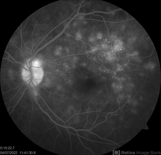
- Condition/keywords
- acute posterior multifocal placoid pigment epitheliopathy (APMPPE), FFA
- Photographer
- jaideep sharma jaipur calgary eye hospital rajasthan india
- Imaging device
- Fundus camera
- Description
- A 50-year old woman presented to us with unilateral progressive and painless visual blurring. She was diagnosed as a case of CSCR and started on topical dorzolamide with no improvement in VA. Her best-corrected visual acuity (BCVA) was RE 6/6 and LE 6/60 . Eye examination revealed vitritis (grade1) with optic disc hyperemia and multiple serous retinal detachments with choroidal striae in the left eye and a normal right eye. She is k/c/o diabetes. Her past ocular and drug histories were unremarkable. Retinal imaging revealed characteristic features of APMPPE in the left eye. All laboratory testing results were inconclusive. VA and OCT findings significantly improved following the treatment with LE posterior sub tenon’s triamcinolone (40 mg/ml). 1 month post injection VA of the left eye reached 6/6 with resolved serous retinal detachments in this eye. This case is unique as it was managed via PST injection rather than conventional steroid therapy


