-
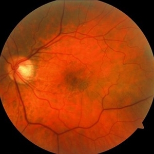 Epiretinal Membrane
May 14 2022 by Rinat Sutiushev
Female, born in 1961. Complains of decreased vision and distortion when reading text. The ocular fundus showed retinal surface wrinkling due to membrane contracture.
Photographer: Rinat Sutiushev
Condition/keywords: Cellophane Maculopathy, epiretinal membrane (ERM), macular pucker
-
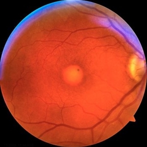 Adult-onset foveomacular vitelliform dystrophy
May 26 2022 by Rinat Sutiushev
Woman born in 1946. Concerns about decreased vision in the right eye, distortions when reading. The ocular fundus of both eyes shows round yellowish deposits (vitelliform material deposits) in the fovea.
Photographer: Rinat Sutiushev
Condition/keywords: adult vitelliform dystrophy, vitelliform lesion, vitelliform macular dystrophy
-
 Adult-onset foveomacular vitelliform dystrophy
May 26 2022 by Rinat Sutiushev
Woman born in 1946. Concerns about decreased vision in right eye, distortions when reading. The ocular fundus of both eyes shows round yellowish deposits (vitelliform material deposits) in the fovea. When autofluorescence photography is performed, hyperautofluorescence is detected.
Photographer: Rinat Sutiushev
Condition/keywords: adult vitelliform dystrophy, vitelliform lesion, vitelliform macular dystrophy
-
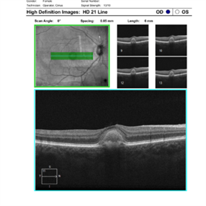 Adult-onset foveomacular vitelliform dystrophy
May 26 2022 by Rinat Sutiushev
Patient born in 1946. Concerns about decreased vision in right eye, distortions when reading. The ocular fundus of both eyes shows round yellowish deposits (vitelliform material deposits) in the fovea. Autofluorescence photography reveals hyperautofluorescence. OCT demonstrates the presence of vitelliform material under the sensitive retina and over the retinal pigment epithelium.
Photographer: Rinat Sutiushev
Condition/keywords: adult vitelliform dystrophy, vitelliform lesion, vitelliform macular dystrophy
-
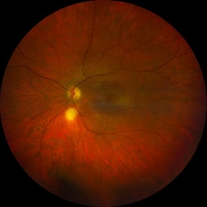
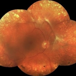 Proliferative diabetic retinopathy
Jul 15 2022 by Rinat Sutiushev
Proliferative diabetic retinopathy with vitreoretinal traction and traction retinal detachment.
Photographer: Rinat Sutiushev
Condition/keywords: proliferative diabetic retinopathy (PDR)
-
 Focal Scleral Nodule
Aug 20 2023 by Rinat Sutiushev
A 48-year-old man with a focal scleral nodule.
Photographer: Rinat Sutiushev, Ophthalmology Center “Zrenie” St. Petersburg
Imaging device: Clarus 500
Condition/keywords: retina, scleral nodule
-
 Focal Scleral Nodule
Aug 20 2023 by Rinat Sutiushev
A 48-year-old man with a focal scleral nodule.
Photographer: Rinat Sutiushev, Ophthalmology Center “Zrenie” St. Petersburg
Condition/keywords: focal scleral nodule, retina
-
 Retinal Astrocytic Hamartoma
Feb 5 2025 by Rinat Sutiushev
Fundus photograph of a 42-year-old man with retinal astrocytic hamartoma type 3.
Photographer: Rinat Sutiushev, Ophthalmological center “Vision”, Saint Petersburg
Imaging device: Heidelberg Spectralis
Condition/keywords: retina
-
 Retinal Astrocytic Hamartoma
Feb 5 2025 by Rinat Sutiushev
Fundus photograph of a 42-year-old man with retinal astrocytic hamartoma type 3.
Photographer: Rinat Sutiushev, Ophthalmological center “Vision”, Saint Petersburg
Imaging device: Heidelberg Spectralis
Condition/keywords: retinal astrocytic hamartoma
-
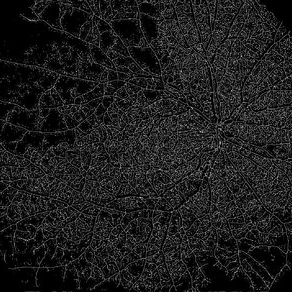 Branch Retinal Vein Occlusion
Apr 10 2025 by Rinat Sutiushev
Ultra-Widefield OCT Angiography of a 77-year-old woman with ischemic occlusion of the superior temporal branch of the central retinal vein with non-proliferative diabetic retinopathy.
Photographer: Rinat Sutiushev, Ophthalmological center “Vision”, Saint Petersburg
Imaging device: TOWARDPI BMIZAR – 400KHZ FULL RANGE SS-OCTA
Condition/keywords: branch retinal vein occlusion (BRVO), nonproliferative diabetic retinopathy, retina
-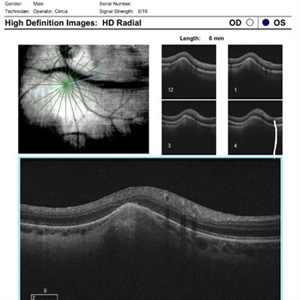
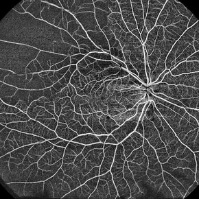
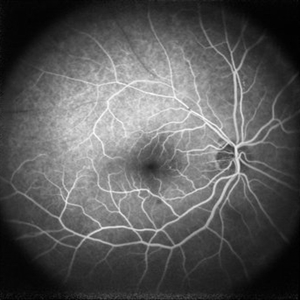 Branch Retinal Vein Occlusion
Apr 10 2025 by Rinat Sutiushev
Fluorescein angiography of a 77-year-old woman with ischemic occlusion of the superior temporal branch of the central retinal vein with non-proliferative diabetic retinopathy.
Photographer: Rinat Sutiushev, Ophthalmological center “Vision”, Saint Petersburg
Imaging device: Heidelberg Spectralis
Condition/keywords: branch retinal vein occlusion (BRVO), nonproliferative diabetic retinopathy
A project from the American Society of Retina Specialists