 Initializing download.
-
 By Deepak Bhojwani, MS
- Uploaded on Mar 15, 2021.
- Last modified by Caroline Bozell on Mar 16, 2021.
- Rating
- Appears in
- 15-Mar-2021
- Condition/keywords
- melanocytoma, optic disc, optical coherence tomography (OCT), enface imaging
- Photographer
- DEEPAK BHOJWANI; OCCURA EYE CARE & RESEARCH CENTER
- Imaging device
-
Fundus camera
OCT - Description
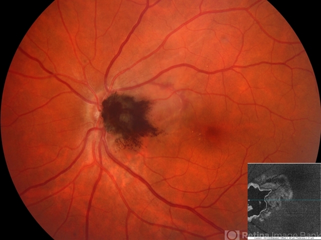
- Fundus photograph of a 49- year-old gentlemen with a characteristic dark brown elevated pigmented mass lesion centered on optic disc and extending into temporal peripapillary area classically suggestive of optic disc melanocytoma. Also note the pigment dispersion and retinal edema just superotemporal to the lesion secondary to tumor necrosis. Inset -Enface OCT image segmented at IS-OS ellipsoid zone level delineating exact horizontal & vertical extent of this tumor mass. Enface OCT imaging also helps in detailing the choroidal extension of such tumors.

")


")
")
")


