-
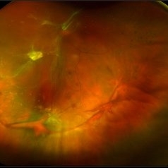 Tractional Retinal Detachment
Aug 22 2019 by Stacie Neview
Ultra-wide field pseudo-color photograph of 47-year-old male with diabetic retinopathy and subsequent diabetic tractional detachment. Patient was lost to follow up for 9 months after receiving anti-VEGF injections with mild PRP and presented with blurry vision.
Photographer: Stacie Neview
Imaging device: Optos
Condition/keywords: diabetic macular edema, diabetic retinopathy, inferior retina, left eye, pan-retinal photocoagulation (PRP), proliferative diabetic retinopathy (PDR), tractional retinal detachment
-
 Subretinal Hemorrhage
Jan 7 2020 by Stacie Neview
Optos fundus photograph of a 74-year-old male with severe subretinal hemorrhage and exudative retinal detachment secondary to peripheral choroidal neovascular membrane.
Photographer: Stacie Neview, Retina Specialists of Michigan, Grand Rapids Michigan, USA
Imaging device: Optos California
Condition/keywords: exudative retinal detachment, subretinal hemorrhage
-
 Choroidal Hemangioma
Jan 19 2021 by Stacie Neview
Ultra wide field fluorescein angiogram of a 44-year-old male with presumed central serous retinopathy. Based on extended ophthalmoscopy, diagnostic ocular ultrasonography, and retinal imaging, the choroidal tumor is most consistent with a choroidal hemangioma. A circumscribed choroidal hemangioma such as this one is unlikely to be associated with an underlying systemic condition and will be further monitored and assessed for possible treatment.
Photographer: Stacie Neview, COA, OSC
Imaging device: Optos California
Condition/keywords: choroidal hemangioma, early phase, fluorescein angiogram (FA), left eye, Optos, ultra-wide field imaging
A project from the American Society of Retina Specialists