 Initializing download.
-
 By SANDEEP KUMAR
Shroff Eye Centre
Co-author(s): Charu Gupta ,Daraius N Shroff ,Cyrus Shroff - Uploaded on Jun 26, 2020.
- Last modified by SANDEEP KUMAR on Jul 2, 2020.
- Rating
- Appears in
- Miscellaneous
- Condition/keywords
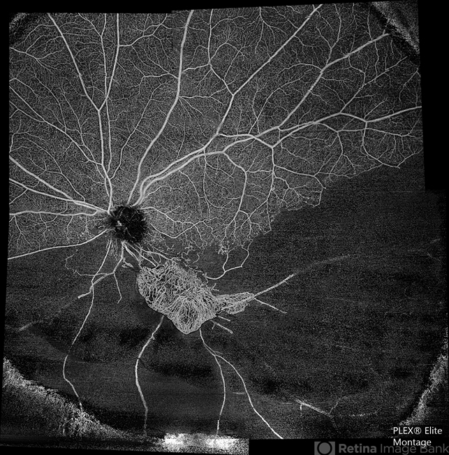
- hemi CRVO
- Photographer
- Sandeep Kumar
- Imaging device
-
Optical coherence tomography system
Optical coherence tomography system Zeiss Plex Elite 9000 - Description
- A 59-year-old man with DM for 18 years operated for mature cataract. Post op left eye had a visual acuity of 20/80. Wide field swept source OCTA revealed gross vessel wipe out in inferior hemi quadrant with branching out neovascular frond inferior to disc with terminal loops, The patient underwent Anti VEGF injection followed by OCTA guided sectoral retinal photocoagulation.Image J software used here to generate reverse image that sharply delineates the non perfusion are


