-
 Two-Lobed Melanotic and Amelanotic Tumor
May 18 2020 by McGill University Health Centre
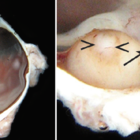
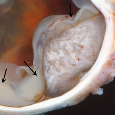
Choroidal melanoma is often asymptomatic and diagnosis is incidental. The tumors may grow beneath the retina, or may break through the Bruch membrane and disrupt the retina. Tumors breaking through the Bruch membrane and disrupting the retina have a characteristic “mushroom” shape. The enucleation specimen in (A) shows a 2-lobed melanotic (arrow) and amelanotic (arrowhead) tumor in the posterior pole of the eye overlying the optic nerve head. In (B), higher magnification shows the optic nerve head and a feeder tumor vessel (arrow). Necrosis is present in the amelanotic tumor (arrowheads).
Condition/keywords: enucleation, tumor
-
 Amelanotic Choroidal Tumor
May 18 2020 by McGill University Health Centre
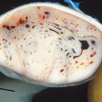
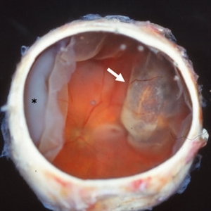
Choroidal melanoma is often asymptomatic and diagnosis is incidental. The tumors may grow beneath the retina, or may break through the Bruch membrane and disrupt the retina. Tumors breaking through the Bruch membrane and disrupting the retina have a characteristic “mushroom” shape. This enucleation specimen shows an amelanotic, dome-shaped choroidal tumor with several dilated blood vessels. The tumor has not infiltrated the sclera, ciliary body, or optic nerve. Note the retinal detachment next to the tumor (arrow).
Condition/keywords: amelanotic, choroidal tumor
-
 Choroidal Tumor
May 18 2020 by McGill University Health Centre
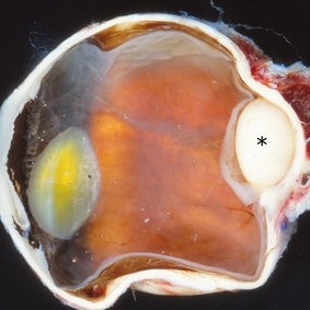
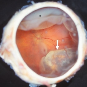
Choroidal melanoma is often asymptomatic and diagnosis is incidental. The tumors may grow beneath the retina, or may break through the Bruch membrane and disrupt the retina. Tumors breaking through the Bruch membrane and disrupting the retina have a characteristic “mushroom” shape. The enucleation specimen in (A) shows a whitish, nodular choroidal tumor at the posterior pole (*). Note the retinal detachment overlying the tumor.
Condition/keywords: choroidal tumor
-
 Choroidal Tumor
May 18 2020 by McGill University Health Centre
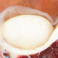
Choroidal melanoma is often asymptomatic and diagnosis is incidental. The tumors may grow beneath the retina, or may break through the Bruch membrane and disrupt the retina. Tumors breaking through the Bruch membrane and disrupting the retina have a characteristic “mushroom” shape. The enucleation specimen in (B) shows a higher magnification of a whitish, nodular choroidal tumor at the posterior pole.
Condition/keywords: choroidal tumor
-
 Pigmented Choroidal Tumor
May 18 2020 by McGill University Health Centre
Choroidal melanoma is often asymptomatic and diagnosis is incidental. The tumors may grow beneath the retina, or may break through the Bruch membrane and disrupt the retina. Tumors breaking through the Bruch membrane and disrupting the retina have a characteristic “mushroom” shape. This enucleation specimen shows a partially pigmented choroidal tumor. The tumor reaches the optic nerve. Note the proteinaceous retinal detachment overlying and adjacent to the tumor (arrows).
Condition/keywords: choroidal tumor
-
 Melanotic Choroidal Tumor
May 18 2020 by McGill University Health Centre
Choroidal melanoma is often asymptomatic and diagnosis is incidental. The tumors may grow beneath the retina, or may break through the Bruch membrane and disrupt the retina. Tumors breaking through the Bruch membrane and disrupting the retina have a characteristic “mushroom” shape. This enucleation specimen shows a melanotic, dome-shaped choroidal tumor (arrow). There is partial retinal detachment with underlying subretinal fluid in the area opposite the tumor (*) and slight macular edema in the posterior pole (seen in the central area of the image).
Condition/keywords: choroidal tumor, enucleation
-
 Melanotic Choroidal Tumor
May 18 2020 by McGill University Health Centre
Choroidal melanoma is often asymptomatic and diagnosis is incidental. The tumors may grow beneath the retina, or may break through the Bruch membrane and disrupt the retina. Tumors breaking through the Bruch membrane and disrupting the retina have a characteristic “mushroom” shape. This enucleation specimen shows a melanotic, dome-shaped choroidal tumor (arrow). There is partial retinal detachment with underlying subretinal fluid in the area opposite the tumor (*) and slight macular edema in the posterior pole (seen in the central area of the image).
Condition/keywords: enucleation
A project from the American Society of Retina Specialists