-
 Choroidal Osteoma
Apr 10 2020 by Dipak Nag, MBBS, FCPS, MSc, FRF
The B scan showed slightly elevated, high reflective choroidal mass (red arrow) with acoustic shadowing of “pseudo-optic nerve” (yellow arrow). The mass persists even in lower gain. A scan showed a high intensity spike.
Photographer: SSN Nishi
Condition/keywords: B scan ultrasound, choroidal osteoma
-
 Old RRD With Retinal Cysts and High Watermark
Apr 10 2020 by Dipak Nag, MBBS, FCPS, MSc, FRF
Intra-operative fundus picture of a 20-year-old boy showing multiple retinal cysts and high watermark in a case of old inferior retinal detachment OD.
Photographer: Dipak
Condition/keywords: high watermark, retinal cyst
-
 CHRRPE
Apr 26 2020 by Dipak Nag, MBBS, FCPS, MSc, FRF
CHRRPE
Condition/keywords: combined hamartoma, retinal pigment epithelium
-
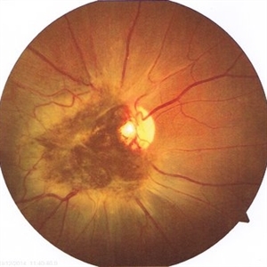
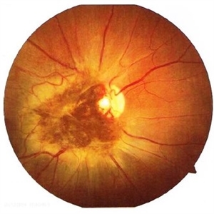 Combined Hamartoma of the Retina and Retinal Pigment Epithelium
Apr 26 2020 by Dipak Nag, MBBS, FCPS, MSc, FRF
A 28-year-old female presented with a deeply pigmented gray- brown, elevated lesion extending from the temporal side of the disc to the macula (OU). Remarkable retinal vasculature with straightening of the distal vessels and dilatation as well as tortuousity of the peri-lesional vessels. The vitreoretinal interface shows gliosis and epi-retinal membrane (ERM) formation.
Photographer: Dipak Nag
Condition/keywords: hamartoma, retinal pigment epithelium
-
 ROP-Zone-I-Stage-3-Plus
Jun 3 2022 by Dipak Nag, MBBS, FCPS, MSc, FRF
Fundus photograph of a child of gestational age 26 weeks and birth weight 1050 grams, shows dilatation and tortuosity of vessels in zone I, extra-retinal fibro-vascular proliferation, hemorrhage with huge peripheral avascular area.
Photographer: Dipak Nag, National Institute of Ophthalmology, Dhaka, Bangladesh
Imaging device: RetCam shuttle
Condition/keywords: retinopathy of prematurity (ROP), retinopathy of prematurity Plus disease, retinopathy of prematurity stage 3, retinopathy of prematurity zone I
A project from the American Society of Retina Specialists