-
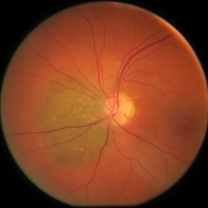 Small, Peripapillary Choroidal Melanoma of the Left Eye - Standard Color
Feb 13 2020 by Michael Seider, MD
Small, peripapillary choroidal melanoma of the left eye. Note the diffuse borders, clumped overlying orange pigment (lipofuscin) and lack of drusen. The standard fundus photograph reveals the true color of the lesion. The wide-field Optos photograph shows the lesion as being more green than the true color, although the lipofuscin remains prominently orange. Wide-field fundus autofluoresence shows a bright signal corresponding to the orange pigment and also superior and inferior to the optic nerve, likely from previous exposure to subretinal fluid. Optical coherence tomography confirms the subretinal fluid seen on examination. B-Scan ultrasonography shows a very low-lying choroidal lesion adjacent to the optic nerve.
-
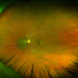 Small, Peripapillary Choroidal Melanoma of the Left Eye - Widefield Color
Feb 13 2020 by Michael Seider, MD
Small, peripapillary choroidal melanoma of the left eye. Note the diffuse borders, clumped overlying orange pigment (lipofuscin) and lack of drusen. The standard fundus photograph reveals the true color of the lesion. The wide-field Optos photograph shows the lesion as being more green than the true color, although the lipofuscin remains prominently orange. Wide-field fundus autofluoresence shows a bright signal corresponding to the orange pigment and also superior and inferior to the optic nerve, likely from previous exposure to subretinal fluid. Optical coherence tomography confirms the subretinal fluid seen on examination. B-Scan ultrasonography shows a very low-lying choroidal lesion adjacent to the optic nerve.
-
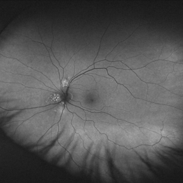 Small, Peripapillary Choroidal Melanoma of the Left Eye - Widefield FAF
Feb 13 2020 by Michael Seider, MD
Small, peripapillary choroidal melanoma of the left eye. Note the diffuse borders, clumped overlying orange pigment (lipofuscin) and lack of drusen. The standard fundus photograph reveals the true color of the lesion. The wide-field Optos photograph shows the lesion as being more green than the true color, although the lipofuscin remains prominently orange. Wide-field fundus autofluoresence shows a bright signal corresponding to the orange pigment and also superior and inferior to the optic nerve, likely from previous exposure to subretinal fluid. Optical coherence tomography confirms the subretinal fluid seen on examination. B-Scan ultrasonography shows a very low-lying choroidal lesion adjacent to the optic nerve.
-
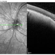 Small, Peripapillary Choroidal Melanoma of the Left Eye - OCT
Feb 13 2020 by Michael Seider, MD
Small, peripapillary choroidal melanoma of the left eye. Note the diffuse borders, clumped overlying orange pigment (lipofuscin) and lack of drusen. The standard fundus photograph reveals the true color of the lesion. The wide-field Optos photograph shows the lesion as being more green than the true color, although the lipofuscin remains prominently orange. Wide-field fundus autofluoresence shows a bright signal corresponding to the orange pigment and also superior and inferior to the optic nerve, likely from previous exposure to subretinal fluid. Optical coherence tomography confirms the subretinal fluid seen on examination. B-Scan ultrasonography shows a very low-lying choroidal lesion adjacent to the optic nerve.
-
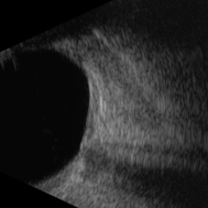 Small, Peripapillary Choroidal Melanoma of the Left Eye - B-Scan
Feb 13 2020 by Michael Seider, MD
Small, peripapillary choroidal melanoma of the left eye. Note the diffuse borders, clumped overlying orange pigment (lipofuscin) and lack of drusen. The standard fundus photograph reveals the true color of the lesion. The wide-field Optos photograph shows the lesion as being more green than the true color, although the lipofuscin remains prominently orange. Wide-field fundus autofluoresence shows a bright signal corresponding to the orange pigment and also superior and inferior to the optic nerve, likely from previous exposure to subretinal fluid. Optical coherence tomography confirms the subretinal fluid seen on examination. B-Scan ultrasonography shows a very low-lying choroidal lesion adjacent to the optic nerve.
A project from the American Society of Retina Specialists