 Initializing download.
-
 By David L Kilpatrick, MD
- Uploaded on Dec 12, 2019.
- Last modified by Caroline Bozell on Dec 12, 2019.
- Rating
- Appears in
- Retinal Capillary Hemangiomas
- Condition/keywords
- retinal capillary hemangioma
- Description
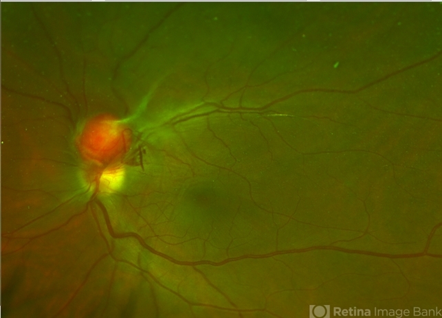
- This is a wide-field color fundus photo showing two distinct retinal capillary hemangiomas. A visually significant epiretinal membrane is also present. Work up with gene testing was negative for VHL. The plan is to proceed with PDT of the two separate lesions (half fluence for the peripapillary lesion), followed by cryotherapy / photocoagulation.


---thumb.jpg/image-square;max$79,0.ImageHandler "Capillay Hemangioma Von Hippel")


---thumb.jpg/image-square;max$79,0.ImageHandler "Retinal Capillary Hemangioma")
---thumb.jpg/image-square;max$79,0.ImageHandler "Retinal Capillary Hemangioma")

