 Initializing download.
-
 By John S. King, MD
Retina Associates, PA - Uploaded on Oct 1, 2019.
- Last modified by Caroline Bozell on Oct 1, 2019.
- Rating
- Appears in
- Miscellaneous
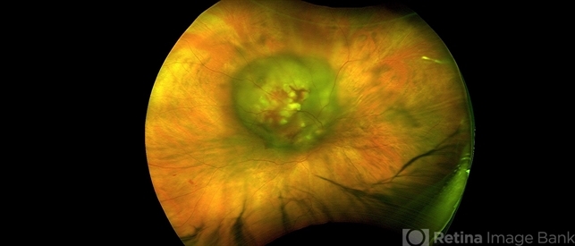
- Condition/keywords
- breast cancer, choroidal metastasis, choroidal lesions
- Photographer
- Kay Dalby
- Imaging device
-
Fundus camera
Optos CA - Description
- 60-year-old white female with four year history of breast cancer associated with metastasis to many organs including the CNS, was sent her to r/o melanoma, found on routine exam. Visual acuity was HM; there was NSC/PSC; there was a unilateral, large choroidal lesion in the posterior pole that was yellow, well circumscribed, with plateau configuration associated with SRF adn heme.


---thumb.jpg/image-square;max$79,0.ImageHandler "Metastatic Breast Cancer")


