 Initializing download.
-
 By Ratimir Lazic, MD, PhD
Eye Clinic Svjetlost
Co-author(s): Marko Lukic, MD - Uploaded on Jan 26, 2013.
- Last modified by Chayal Patel on Jan 29, 2013.
- Reviewed by Chayal Patel
- Rating
- Appears in
- CRVO Treated with Ranibizumab
- Condition/keywords
- central retinal vein occlusion (CRVO)
- Photographer
- Marko Lukic, MD
- Imaging device
-
Fundus camera
Zeis Visucam Lite 2 - Description
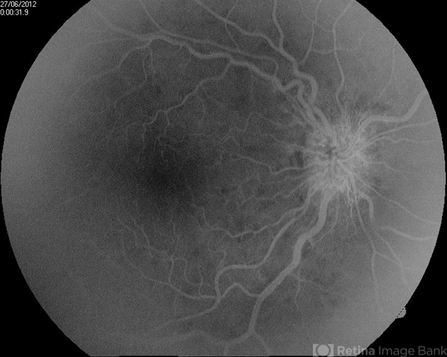
- FAG image of a 67-year-old female after 3 consecutive ranibizumab injections. Invenous phase hyperphlorescence on optic nerve due to presence of optociliary shunts can be seen. No macular edema is present.


