-
 Central Retinal Vein Occlusion - After 3 Consecutive Ranibizumab Injections
Jan 26 2013 by Ratimir Lazic, MD, PhD
Color fundus photography of a 67-year-old female. Intraretinal hemorrhages in posterior pole, tortuous and dilated veins with optociliary shunts visible on optic nerve head. No macular edema can be noticed.
Photographer: Marko Lukic, MD
Imaging device: Zeis Visucam Lite 2
Condition/keywords: central retinal vein occlusion (CRVO)
-
 Central Retinal Vein Occlusion - After 3 Consecutive Ranibizumab Injections
Jan 26 2013 by Ratimir Lazic, MD, PhD
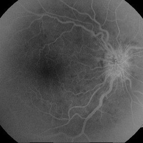
FAG image of a 67-year-old female after 3 consecutive ranibizumab injections. Invenous phase hyperphlorescence on optic nerve due to presence of optociliary shunts can be seen. No macular edema is present.
Photographer: Marko Lukic, MD
Imaging device: Zeis Visucam Lite 2
Condition/keywords: central retinal vein occlusion (CRVO)
-
 Central Retinal Vein Occlusion - After 3 Consecutive Ranibizumab Injections
Jan 26 2013 by Ratimir Lazic, MD, PhD
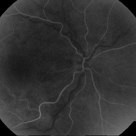
FAG image(arterial phase) of a 67-year-old female after 3 consecutive ranibizumab intravitreal injections due to CME caused by CRVO.
Photographer: Marko Lukic, MD
Imaging device: Zeis Visucam Lite 2
Condition/keywords: central retinal vein occlusion (CRVO)
A project from the American Society of Retina Specialists