 Initializing download.
-
 By Ratimir Lazic, MD, PhD
Eye Clinic Svjetlost
Co-author(s): Marko Lukic, MD - Uploaded on Jan 26, 2013.
- Last modified by Chayal Patel on Jan 29, 2013.
- Reviewed by Chayal Patel
- Rating
- Appears in
- Eals Disease
- Condition/keywords
- Eales disease
- Photographer
- Marko Lukic, MD
- Imaging device
-
Fundus camera
Zeis Visucam Lite 2 - Description
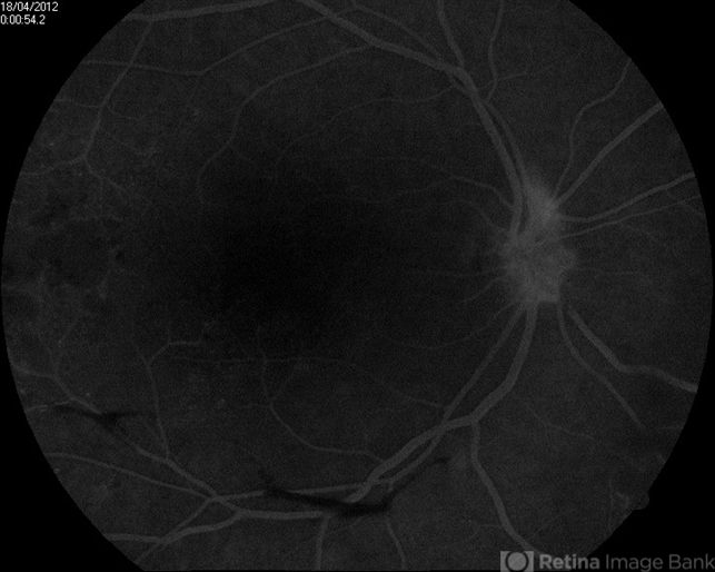
- FAG image of a 28-year-old male. Late venous phase with few important notes can be seen; neovascularisations of the optic disc, retrohyaloid hemorrhage in lower quadrants (hypoflorescent area) and hyperflorescent dots in macula.


