-
 Tuberous Sclerosis OCT Final
Aug 28 2019 by Nisarg Joshi, MD
26-year-old female with history of tuberous sclerosis was found to have retinal phakomatous hamartomas in the right eye (peripapillary pale yellow lesion) and the left eye (opaque lesion in superior macula). This OCT shows the typical homogeneous appearance of these benign retinal lesions. The macula also shows RPE changes in both eyes.
Photographer: Nisarg Joshi, Geisinger Eye Institute, Danville, PA
Imaging device: Heidelberg Spectralis
Condition/keywords: hamartoma, phakoma, tuberous sclerosis
-
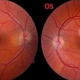 Retinal Phakomatous Hamartoma in Tuberous Sclerosis- Fundus Photos
Aug 28 2019 by Nisarg Joshi, MD
26-year-old female with history of tuberous sclerosis was found to have retinal phakomatous hamartomas in the right eye (peripapillary pale yellow lesion) and the left eye (opaque lesion in superior macula). The macula also shows RPE changes in both eyes.
Photographer: Nisarg Joshi, Geisinger Eye Institute, Danville, PA
Condition/keywords: hamartoma, phakoma, tuberous sclerosis
A project from the American Society of Retina Specialists