 Initializing download.
-
 By Natalie Loyacano, COMT, OCS-R,OSA, ROUB
VitreoRetinal Eye Center
Co-author(s): Avit Gremillion, MD, VitreoRetinal Eye Center, Biloxi MS - Uploaded on Oct 30, 2015.
- Last modified by Caroline Bozell on Oct 30, 2015.
- Rating
- Appears in
- Miscellaneous
- Condition/keywords
- choroidal mass
- Photographer
- Amy Gunter, VitreoRetinal Eye Center, Biloxi MS
- Imaging device
-
Fundus camera
Topcon - Description
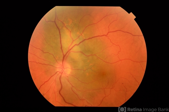
- Fundus photograph of 50 year-old male with a suspicious peripapillary pigmented lesion. Patient sees a spot in his vision that has progressively worsen over the past month.
---thumb.jpg/image-square;max$79,0.ImageHandler "Vitrectomy Choroidal Mass")


