 Initializing download.
-
 By Unnati Vishwanath Shukla, M. S. ,DNB, FVRS FNERF, MNAMS,PhD Scholar(Retina)
Nagpal's Retina Foundation, Ahmedabad
Co-author(s): Tejas Desai . Shaileen Parikh - Uploaded on May 24, 2019.
- Last modified by Caroline Bozell on Aug 2, 2019.
- Rating
- Appears in
- retina
- Condition/keywords
- choroidal tuberculoma, pregnancy, macula lesion, Quantiferon gold, mantoux test
- Photographer
- Unnati Shukla, C.H. Nagri Eye Hospital, NHL medical college, Ahmedabad,Gujarat,India.
- Imaging device
- Fundus camera
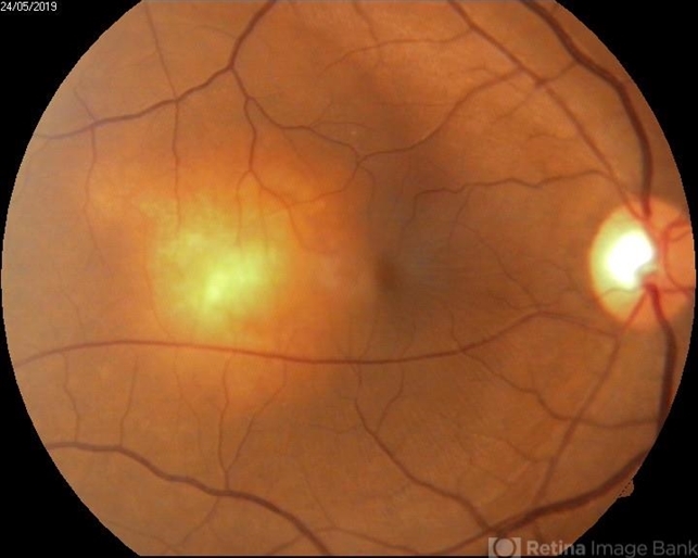
- Description
- A 29-year-old pregnant female patient (in the third trimester of pregnancy) presented with the complaints of blurring of vision in the right eye and metamorphosia since 1 month. On examination a solitary yellowish elevated subretinal mass of about 2 disc diameters lesion was noted in the right eye temporal to the macula with minimal subretinal fluid around the lesion. Left eye findings were normal. Further investigations revealed normal Chest X- ray and abdominal USG and negative sputum AFB analysis. Thorough uveitic profile was done to rule out other infective and immune causes of choroidal lesion . Positive findings revealed positive mantoux test , positive interferon gamma release essay (Quantiferon Gold). Further aqueous tapping was done which showed presence of acid fast bacilli. Final diagnosis of right eye isolated choroidal tuberculoma was made and appropriate treatment was initiated.


---thumb.jpg/image-square;max$79,0.ImageHandler "Age Related Macular Degeneration - Geographic Atrophy")
---thumb.jpg/image-square;max$79,0.ImageHandler "Age Related Macular Degeneration - Geographic Atrophy")
---thumb.jpg/image-square;max$79,0.ImageHandler "Age Related Macular Degeneration - Geographic Atrophy")
---thumb.jpg/image-square;max$79,0.ImageHandler "Age Related Macular Degeneration")


