-
 Posterior Uveitis
Apr 8 2019 by Gary R. Cook, MD, FACS
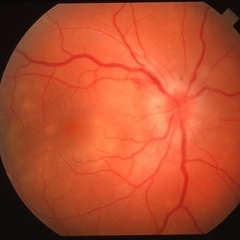
37-year-old white male with mild vitritis, optic disc hyperemia and edema, peripapillary hemorrhages and yellow-white spots in temporal macula OD; V.A. = 20/30.
Imaging device: Topcon VT-50
Condition/keywords: posterior uveitis
-
 Posterior Uveitis
Apr 8 2019 by Gary R. Cook, MD, FACS
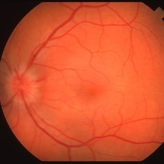
37-year-old white male with mild vitritis, optic disc hyperemia and edema, and a couple of peripapillary NFL hemorrhages OS; V.A. = 20/20-1
Imaging device: Topcon VT-50
Condition/keywords: posterior uveitis
-
 Posterior Uveitis
Apr 8 2019 by Gary R. Cook, MD, FACS
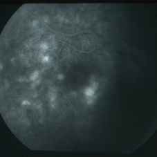
Mid-phase (64 seconds) fluorescein angiogram image showing mild leakage and early staining of the yellow-white spots in the temporal macula of the right eye; V.A. = 20/30.
Imaging device: Topcon VT-50
Condition/keywords: FA mid phase, posterior uveitis
-
 Posterior Uveitis
Apr 8 2019 by Gary R. Cook, MD, FACS
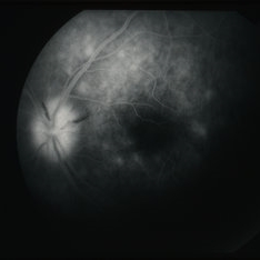
Late-phase (219 seconds) fluorescein angiogram image of the left eye showing late staining of the optic disc and of numerous spots deep to the retina; also blocked fluorescence from the 2 NFL hemorrhages on the optic disc; V.A. = 20/20-1
Imaging device: Topcon VT-50
Condition/keywords: FA late phase, posterior uveitis
A project from the American Society of Retina Specialists