 Initializing download.
-
 By Fawwaz F Al Mamoori, MD, Medical Retina Consultant
Sharif Eye Center - Uploaded on Dec 21, 2018.
- Last modified by Caroline Bozell on Dec 21, 2018.
- Rating
- Appears in
- 21-Dec-2018
- Condition/keywords
- optical coherence tomography (OCT), paracentral acute middle maculopathy
- Photographer
- Dr.Fawwaz Al Mamoori (Al Mamoori Eye Clinic)
- Imaging device
-
Optical coherence tomography system
Triton Swept Source OCT (TOPCON) - Description
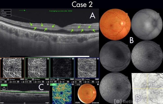
- 70-year-old female patient known to have hypertension, presented with acute deterioration of left eye vision, best corrected visual acuity was 6/60. Fundoscopic exam showed abnormal foveal reflex whiting .SS-OCT B scan showed also a hypereflectivity of the inner plexiform layer (IPL), inner nuclear layer (INL) and OPL layer(figure-2, A). FA images were also normal(figure-2 B). Segmented angiographic images elucidate ischemia and capillary drop out predominantly at the level of DCP but less severe than Case 1 (fig 2, C). Correspondingly, Enface highlights hyper reflective areas in a fern like distribution in the middle retina at similar depth of ischemic lesions demonstrated on B scans and OCTA (fig 2, D)

")


")
")
")


