-
 Cone-Rod Dystrophy
Jun 23 2018 by Hossein Ameri, MD, PhD, FRCSI, MRCOphth
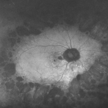
Ultra-wide field autofluorescence of a patient with cone-rod dystrophy showing mid peripheral ring of hypo autofluorescence, as well as autofluorescence changes in the macular area.
Imaging device: Optos
Condition/keywords: cone dystrophy
-
 Cone-Rod Dystrophy
Jun 23 2018 by Hossein Ameri, MD, PhD, FRCSI, MRCOphth
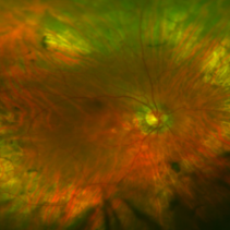
Ultra-wide field fundus photo of a patient with cone-rod dystrophy showing peripheral pigment clumping, mid-peripheral retinal pigment epithelial atrophy, and pigmentary changes in the macula.
Imaging device: Optos
Condition/keywords: cone dystrophy
A project from the American Society of Retina Specialists