File number: 28296
Comments
-
 Hosam Attia, MD (June 26 2018)
Nicely Captured!
Thank you for sharing and for the description, since PFCL could be easily missed without that.
was there a history of trauma/ zonulysis , specially w/ PCFL in the A.C in a phakic patient ?
why the PFCL is not settling completely inferiorly in the A.C/ Angle, while patient is upright for photography , specially with the surface tension exerted on it, superiorly form the gas bubble, which is another reason for it to settle down even more !
Is the angle shallow inferiorly ??
Is there corneal touch as well ??
Was Viscoelastic used intra-operatively in the AC for any reason and may be retained, but if it is two weeks, it should have washed out any ways !!
Sign in to comment.
 Initializing download.
-
 By Maria Stephanie R. Jardeleza, MD
North Carolina Retina Associates
Co-author(s): M. Stephanie R. Jardeleza, M.D., Retina Specialist, San Antonio Eye Center, SA, TX - Uploaded on Jun 21, 2018.
- Last modified by Caroline Bozell on Jun 22, 2018.
- Rating
- Appears in
- Miscellaneous
- Condition/keywords
- vitreous substitutes, retained perfluorocarbon
- Photographer
- Andy Zepeda, COA, Retina Clinic, San Antonio Eye Center, San Antonio, TX
- Imaging device
- Photo slit lamp biomicroscope
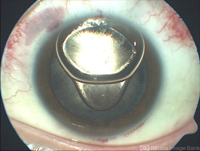
- Description
- Anterior segment photographs of 30-year-old male who underwent superior rhegmatogenous retinal detachment repair with intraocular gas tamponade. Perfluorocarbon was used to flatten the macula to prevent a macular fold and was removed during PFC/air exchange. Post operative week two visit shows gas migration into the anterior chamber with retained PFC on the posterior aspect of the gas bubble/anterior surface of the lens. Patient had been maintaining face down positioning.


