Patients with Diabetic Retinopathy
-
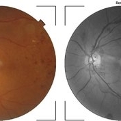
 Diabetic Retinopathy
Apr 5 2018 by JYOTI PATIL, Ph.D.
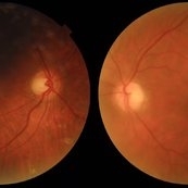
Scattered hemorrhages in the left eye. Fundus photograph of a 45-year-old man with diabetic retinopathy.
Photographer: Dr.Aditya Kelkar
Condition/keywords: diabetic retinopathy
-
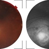
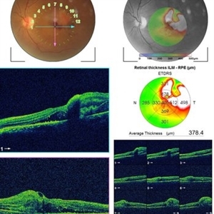
 Diabetic Retinopathy
Apr 5 2018 by JYOTI PATIL, Ph.D.
OS(L) Macula. Fundus photograph of a 45-year-old man with a Diabetic Retinopathy, observed with information of Retinal thickness.
Photographer: Dr.Aditya Kelkar
Condition/keywords: diabetic retinopathy
-
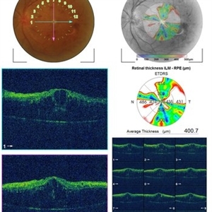
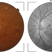
 Diabetic Retinopathy
Apr 5 2018 by JYOTI PATIL, Ph.D.
Fundus photographs of diabetic retinopathy eye observed with change in optic axis.
Photographer: Dr.Aditya Kelkar
Condition/keywords: diabetic retinopathy
-
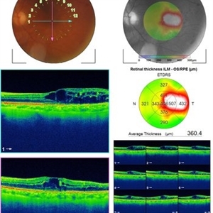 Diabetic Retinopathy
Apr 5 2018 by JYOTI PATIL, Ph.D.
OS(L) Macula. Fundus photograph of an 45-year-old man with diabetic retinopathy, observed with information of retinal thickness.
Photographer: Dr.Aditya Kelkar
Condition/keywords: diabetic retinopathy
-
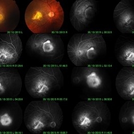
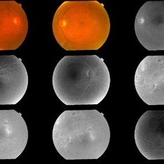
 Neovascularization of the Disc
Apr 5 2018 by JYOTI PATIL, Ph.D.
OS(L).Color fundus photography of a 59-year-old male one week after intravitreal bevacizumab was injected. We can see the effect on the neovascularizations of the disc where their appearance is significant less compared with previous image.
Photographer: Dr.Aditya Kelkar
Condition/keywords: diabetic retinopathy, neovascularization (NV), neovascularization of the disc (NVD)
-
 Neovascularization of the Disc
Apr 5 2018 by JYOTI PATIL, Ph.D.
Color fundus photography of a 62-year-old male one week after intravitreal bevacizumab was injected. We can see the effect on the neovascularizations of the disc where their appearance is significant less compared with previous image.
Photographer: Dr.Aditya Kelkar
Condition/keywords: diabetic retinopathy, neovascularization of the disc (NVD)
-
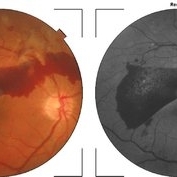
 Diabetic Retinopathy
Apr 5 2018 by JYOTI PATIL, Ph.D.
Fundus photograph with diabetic retinopathy observed with hemorrhage.
Photographer: Dr.Aditya Kelkar
Condition/keywords: diabetic retinopathy
-
 Diabetic Retinopathy
Apr 5 2018 by JYOTI PATIL, Ph.D.
Boat-shaped hemorrhage in a patient with retro-hyaloid hemorrhage associated with proliferative diabetic retinopathy.
Photographer: Dr.Aditya Kelkar
Condition/keywords: diabetic retinopathy
-
 Diabetic Retinopathy
Apr 5 2018 by JYOTI PATIL, Ph.D.
A FAG image of a 74-year-old female. Diabetic changes of the posterior pole and midperipheral retina can be seen. dots (microaneurisms) and hypoflorescent areas (intraretinal hemorrhages) can be seen.
Photographer: Dr.Aditya Kelkar
Condition/keywords: diabetic retinopathy
-
 Neovascularizations of the Disc
Apr 5 2018 by JYOTI PATIL, Ph.D.
Color fundus photography of a 54-year-old male one week after intravitreal bevacizumab was injected. We can see the effect on the neovascularizations of the disc where their appearance is significant less compared with previous image.
Photographer: Dr.Aditya Kelkar
Condition/keywords: diabetic retinopathy
-
 Bilateral Dengue Retinitis
Apr 5 2018 by JYOTI PATIL, Ph.D.
Right eye of a 40-year-old lady recovering from bilateral dengue retinitis shows dengue foveolitis, neovascularisation disc (NVD) and vitreous hemorrhage.
Photographer: Dr.Aditya Kelkar
Condition/keywords: bilateral dengue retinitis, diabetic retinopathy, vitreous hemorrhage
-
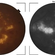 Diabetic Retinopathy
Apr 5 2018 by JYOTI PATIL, Ph.D.
Fundus photograph of a 45-year-old man with diabetic retinopathy having severe cotton wool spot.
Photographer: Dr.Aditya Kelkar
Condition/keywords: diabetic retinopathy