 Initializing download.
-
 By Dhaivat Shah
Sankara Nethralaya - Uploaded on Mar 31, 2018.
- Last modified by Caroline Bozell on Apr 3, 2018.
- Rating
- Appears in
- Imaging marvels
- Condition/keywords
- cystoid macular edema (CME), multicolor
- Photographer
- Dr Dhaivat Shah
- Imaging device
- Heidelberg Spectralis
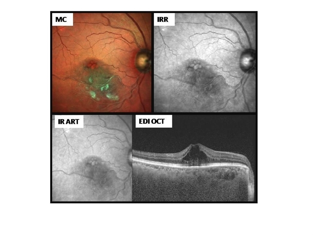
- Description
- This is a case of a 50-year-old female presenting with right eye IT BRVO with ME. Unarguably, the first modality of investigation would be an OCT along with a fundus photograph. Instead of using a conventional fundus photo, a multicolor image was captured. It beautifully highlighted the cystic pattern of macular edema, hemorrhages and sclerosed vessel, which corresponded well with the OCT findings. Multi-color scanning laser imaging uses three laser wavelengths simultaneously to provide diagnostic images that show distinct structures at different depths within the retina. The high-resolution, detailed MultiColor images can highlight structures and pathologies not visible on ophthalmoscopy and fundus photography. This imaging modality has the potential to replace conventional color fundus photography in the near future.
")
")
")
")
")
")


---thumb.jpg/image-square;max$79,0.ImageHandler "Intermediate Uveiris and CME")
---thumb.jpg/image-square;max$79,0.ImageHandler "Intermediate Uveitis and CME")