 Initializing download.
-
 By P. Mahesh Shanmugam, MBBS, DO, FRCSEd, PhD, FAICO
Sankara Eye Hospitals
Co-author(s): Dr Rajesh Ramanjulu - Uploaded on Dec 28, 2015.
- Last modified by Caroline Bozell on Jan 3, 2016.
- Rating
- Appears in
- Miscellaneous
- Condition/keywords
- choroidal osteoma
- Imaging device
- Fundus camera
- Description
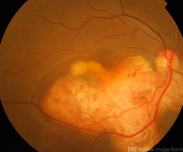
- A 28-year-old female with typical choroidal osteoma as yellow subretinal minimally elevated lesion with scalloped margin. peripheral part of the lesion is orange in color, the older central part yellow in color.


---thumb.jpg/image-square;max$79,0.ImageHandler "Choroidal osteoma case 1 no 2")
---thumb.jpg/image-square;max$79,0.ImageHandler "Choroidal osteoma case 1 no 3")