 Initializing download.
-
 By Marc C. Peden, MD
Retina Associates of Florida
Co-author(s): Marc Peden, M.D., Ivan Suner M.D., Mark Hammer M.D. - Uploaded on Sep 17, 2015.
- Last modified by Caroline Bozell on Sep 17, 2015.
- Rating
- Appears in
- Miscellaneous
- Condition/keywords
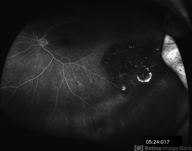
- adenocarcinoma arising from CHRPE
- Photographer
- Janet Traynom COT
- Imaging device
-
Scanning laser ophthalmoscope
Optos P200MA - Description
- 49-year-old female referred for presumed ocular melanoma. On examination was noted to have darkly pigmented lesion in the temporal retina of left eye. Lesion had characteristic scalloped edges with central lacunae, however, on ultrasonography was noted to have 1.8mm of elevation with high internal reflectivity. IVFA shows absence of dual circulation with areas of window defect. Findings were consistent with those described by Shields et al., in their April 2001 article in Archives of Ophthalmology.
