An 83 year-woman returned from wintering in Florida after having been treated with intravitreal antiVEGF OD for exudative AMD. The visual acuity measured 20/200. An RPE tear was noted. Note the scrolled and redundant RPE just temporal to the fovea and the absent RPE more temporally. OCT revealed the RPE tear with some scolled redundant RPE more centrally.
-
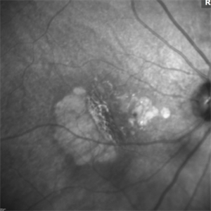 RPE Tear: Infrared Photo
May 2 2015 by Thomas A. Ciulla, MD, MBA, FASRS
Infrared image: Note the scrolled and redundant RPE just temporal to the fovea and the absent RPE more temporally where there are visible larger choroidal vessels.
Photographer: Stuart Alfred
Condition/keywords: choroidal neovascular membrane (CNVM), retinal pigment epithelium (RPE) tear, wet age-related macular degeneration (wet AMD)
-
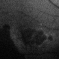 RPE Tear: Autofluorescence
May 2 2015 by Thomas A. Ciulla, MD, MBA, FASRS
Autofluorescence image: Note the discrete hypo-autofluorescence due to the absent RPE temporally. Note also the hypo-autofluorescence due to geographic atrophy centrally.
Photographer: Stuart Alfred
Condition/keywords: choroidal neovascular membrane (CNVM), retinal pigment epithelium (RPE) tear, wet age-related macular degeneration (wet AMD)
-
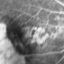 RPE Tear: Fluorescein Angiography
May 2 2015 by Thomas A. Ciulla, MD, MBA, FASRS
Mid Phase Fluorescein Angiogram: The scrolled and redundant RPE just temporal to the fovea blocks underlying choroidal fluorescence. The absent RPE, more temporally, results in a window defect with intense hyperfluorescence.
Photographer: Stuart Alfred
Condition/keywords: choroidal neovascular membrane (CNVM), retinal pigment epithelium (RPE) tear, wet age-related macular degeneration (wet AMD)
-
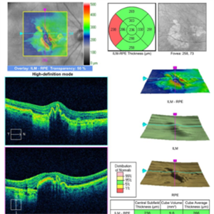 RPE Tear: OCT
May 2 2015 by Thomas A. Ciulla, MD, MBA, FASRS
OCT revealed the RPE tear with some scolled redundant RPE more centrally.
Condition/keywords: choroidal neovascular membrane (CNVM), retinal pigment epithelium (RPE) tear, wet age-related macular degeneration (wet AMD)