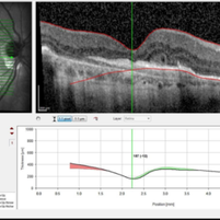
This 85 year-old man has a history of exudative AMD on the right and has undergone numerous antiVEGF injections over the past 8 years. Visual acuity measured 20/60. OCT shows subretinal hyperreflective material with overlying outer retinal tubulation (round or oval structures with hyperreflective borders). Infrared, red-free, autofluorescence and fluorescein angiogram images reveal geographic atrophy and no active choroidal neovascular membrane. Outer retinal tubulation is thought to result from invagination of photoreceptors at the junction of intact and atrophic outer retina. As in this case, greater lesion size, subretinal hyperreflective material and geographic atrophy are all associated with outer retinal tubulation. Poor visual function is also associated with outer retinal tubulation. Some reports suggest that outer retinal tubulation may be a predictor of enlargement of geographic atrophy.
-
 Outer Retinal Tubulation
Jan 10 2015 by Thomas A. Ciulla, MD, MBA, FASRS
This 85-year-old man has a history of exudative AMD on the right and has undergone numerous anti-VEGF injections over the past 8 years. Visual acuity measured 20/60. OCT shows subretinal hyperreflective material with overlying outer retinal tubulation (round or oval structures with hyperreflective borders). Outer retinal tubulation is thought to result from invagination of photoreceptors at the junction of intact and atrophic outer retina. As in this case, greater lesion size, subretinal hyperreflective material and geographic atrophy are all associated with outer retinal tubulation. Poor visual function is also associated with outer retinal tubulation. Some reports suggest that outer retinal tubulation may be a predictor of enlargement of geographic atrophy.
Photographer: Stuart Alfred
Condition/keywords: outer retinal tubulation
-
 Outer Retinal Tubulation
Jan 10 2015 by Thomas A. Ciulla, MD, MBA, FASRS
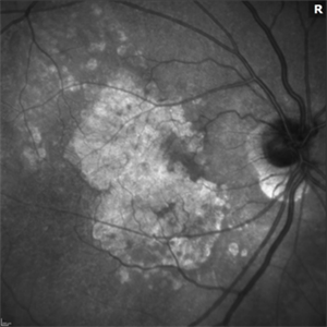
Infrared images reveal geographic atrophy.
Photographer: Stuart Alfred
Condition/keywords: outer retinal tubulation
-
 Outer Retinal Tubulation
Jan 10 2015 by Thomas A. Ciulla, MD, MBA, FASRS
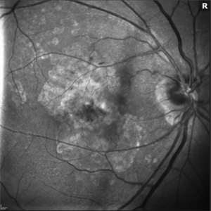
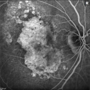
Red-free images reveal geographic atrophy.
Photographer: Stuart Alfred
Condition/keywords: outer retinal tubulation
-
 Outer Retinal Tubulation
Jan 10 2015 by Thomas A. Ciulla, MD, MBA, FASRS
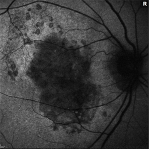
Autofluorescence images reveal geographic atrophy .
Photographer: Stuart Alfred
Condition/keywords: outer retinal tubulation
-
 Outer Retinal Tubulation
Jan 10 2015 by Thomas A. Ciulla, MD, MBA, FASRS
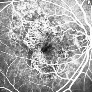
Fluorescein angiogram images reveal geographic atrophy and no active CNVM currently.
Photographer: Stuart Alfred
Condition/keywords: outer retinal tubulation
-
 Outer Retinal Tubulation
Jan 10 2015 by Thomas A. Ciulla, MD, MBA, FASRS
Fluorescein angiogram images reveal geographic atrophy and no active CNVM currently.
Photographer: Stuart Alfred
Condition/keywords: outer retinal tubulation